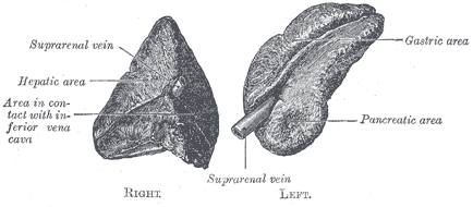
Gray's Fig. 1183 – Suprarenal glands viewed from the front.
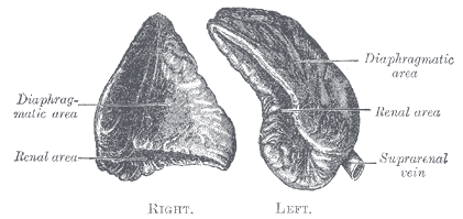
Gray's Fig. 1184 – Suprarenal glands viewed from behind.
In mammals, the adrenal glands (also known as suprarenal glands or colloquially as kidney hats) are the triangle-shaped endocrine glands that sit atop the kidneys; their name indicates that position (ad, "near" or "at" + renes, "kidneys"). They are chiefly responsible for regulating the stress response through the synthesis of corticosteroids and catecholamines, including cortisol and adrenaline.
Overview[]

Above each human kidney is one of the two adrenal glands.

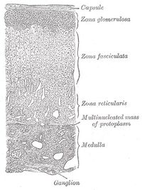
Layers of the adrenal cortex
Anatomically, the adrenal glands are located in the abdomen, situated on the anteriosuperior aspect of the kidneys. In humans, the adrenal glands are found at the level of the 12th thoracic vertebra and receive their blood supply from the adrenal arteries.
It is separated into two distinct structures, the adrenal medulla and the adrenal cortex, both of which receive regulatory input from the nervous system. As its name suggests, the adrenal medulla is at the center of the adrenal gland surrounded by the adrenal cortex.
The adrenal medulla is the body's main source of the catecholamine hormones epinephrine and norepinephrine. By contrast, some cells of the adrenal cortex belong to the hypothalamic-pituitary-adrenal axis and are the source of cortisol synthesis. Other cortical cells produce androgens such as testosterone, while some regulate water and electrolyte concentrations by secreting aldosterone.
Adrenal medulla[]
Composed mainly of hormone-producing chromaffin cells, the adrenal medulla is the principal site of the conversion of the amino acid tyrosine into the catecholamines epinephrine and norepinephrine (also called adrenaline and noradrenaline, respectively). Medullary cells are derived from the embryonic neural crest and, as such, are simply modified neurons. In particular, they are modified postganglionic cells of the sympathetic nervous system that have lost their axons and dendrites, receiving innervation from corresponding preganglionic fibers. Moreover, as the synapses between pre- and postganglionic fibers are called ganglia, the adrenal medulla is actually a ganglion of the sympathetic nervous system.
In response to stressors such as exercise or imminent danger, medullary cells release catecholamines into the blood in a 70:30 ratio of epinephrine to norepinephrine. Notable effects of epinephrine and norepinephrine include increased heart rate, blood vessel constriction, bronchiole dilation, and increased metabolism, all of which are characteristic of the fight-or-flight response.
Adrenal cortex[]
Situated along the perimeter of the adrenal gland, the adrenal cortex mediates the stress response through the production of mineralocorticoids and glucocorticoids, including aldosterone and cortisol respectively. It is also a secondary site of androgen synthesis.
The cortex can be divided into three distinct layers of tissue based on their organization. The most superficial cortical layer is the zona glomerulosa, which produces mineralocorticoids (eg, aldosterone). Beneath the glomerulosa are the zonae fasciculata and reticularis, which produce glucocorticoids (eg, cortisol) and weak androgens (eg, dehydroepiandrosterone).
All adrenocortical hormones are synthesised from cholesterol. Cholesterol is transported into the inner mitochondrial membrane by steroidogenic acute regulatory protein (StAR), where it is converted into pregnenolone by the enzyme CYP11A. Accordingly, production of hormones in all three layers of the adrenal cortex is limited by the transportation of cholesterol into the mitochondria and by its conversion into pregnenolone. Pregnenolone can be either dehydrogenated to progesterone or hydroxylated to 17-alpha-hydroxypregnenolone. The steps up to this point occur in many steroid-producing tissues. Subsequent steps, however, only occur in the adrenal cortex.
- Progesterone ->(hydroxylation at C21)-> Deoxycorticosterone ->(two further hydroxylations)-> Aldosterone
- Progesterone -> (hydroxylation at C17)-> 17-alpha-hydroxyprogesterone ->(hydroxylation)-> Deoxycortisol ->(hydroxylation)-> Cortisol
Zona glomerulosa[]
The zona glomerulosa is the most superficial layer of the adrenal cortex, lying directly beneath the adrenal gland's capsule. Its cells are arranged in spherical clusters (glomus is Latin for "ball").
In response to increased potassium levels or decreased blood flow to the kidneys, cells of the zona glomerulosa secrete the mineralocorticoid aldosterone into the blood as part of the renin-angiotensin system. Aldosterone regulates the body's concentration of electrolytes, primarily sodium and potassium, by acting on the distal convoluted tubule of kidney nephrons to:
- increase sodium reabsorption
- increase potassium excretion
- increase water reabsorption through osmosis
Zona fasciculata[]
Cells of the zona fasciculata sit directly beneath the zona glomerulosa and are organized in bundles (or fascicles). The zona chiefly produces glucocorticoids (eg, cortisol) and a small amount of weak androgens (eg, dehydroepiandrosterone).
Cortical cells responsible for the production of glucocorticoids are the primary effectors of adrenocorticotropic hormone (ACTH). The hypothalamus secretes corticotropin-releasing hormone which stimulates the anterior pituitary gland to release ACTH; another hypothalamic hormone, arginine vasopressin augments ACTH secretion, with the two together stimulating larger release than ACTH in isolation. ACTH acts on the adrenal cortex to stimulate the release of glucocorticoids. This three-organ endocrine system is commonly called the hypothalamic-pituitary-adrenal axis.
The primary glucocorticoid released by the adrenal gland is cortisol. Upon binding to its target, cortisol enhances metabolism in several ways:
- stimulating the release of amino acids from the body
- stimulating lipolysis, the breakdown of fat
- stimulating gluconeogenesis, the production of glucose from newly-released amino acids and lipids
- conserving glucose by inhibiting uptake into muscle and fat cells
In addition to glucocorticoid production, the fasciculata is a secondary source of androgens such as testosterone, dihydrotestosterone (DHT), androstenedione, and dehydroepiandrosterone (DHEA). These enhance muscle mass, stimulate cell growth, and aid in the development of the secondary sex characteristics.
Zona reticularis[]
The innermost layer of the adrenal cortex, the zona reticularis sits beneath the zona fasciculata and atop the adrenal medulla. Its cells are arranged in a network of cords (a reticulum) and have the same functions as cells of the zona fasciculata. It is thought that the zona reticularis is the principal source of glucocorticoids and adrenal androgens, with the zona fasciculata activating only after prolonged stimulation.
Pathology[]
Adrenal cortex:
- Hypoadrenalism (e.g. due to Addison's disease)
- Cushing's syndrome
- Congenital adrenal hyperplasia
- Conn's syndrome
Adrenal medulla:
- Pheochromocytoma is a catecholamine-secreting tumor of the adrenal medulla.
Blood supply[]
Although variations of the blood supply to the adrenal glands (and indeed the kidneys themselves) are common, there are usually three arteries that supply each adrenal gland: the superior, middle and inferior suprarenal (or adrenal) arteries.
Each (left and right) superior suprarenal artery is a branch of the phrenic artery on that side of the body. The left and right phrenic arteries supply the diaphragm, and come off the aorta.
The middle suprarenal artery is a branch directly off the aorta, while the inferior suprarenal artery branches from the aorta or the renal artery.
See also[]
References[]
- Adrenal Gland, from Colorado State University
- MedlinePlus Medical Encyclopedia: Adrenal Glands
Human anatomy, endocrine system: endocrine glands | |||||||||||||||||||||||||
|---|---|---|---|---|---|---|---|---|---|---|---|---|---|---|---|---|---|---|---|---|---|---|---|---|---|
| Hypothalamic/ pituitary axes |
| ||||||||||||||||||||||||
| Pineal gland |
Pinealocyte · Corpora arenacea | ||||||||||||||||||||||||
| Islets of pancreas |
Alpha cell · Beta cell · Delta cell · PP cell · Epsilon cell | ||||||||||||||||||||||||
de:Nebenniere es:Glándula suprarrenal fr:Glande surrénale lt:Antinkstis nl:Bijnier no:Binyre pt:Glândula supra-renal sv:Binjure
| This page uses Creative Commons Licensed content from Wikipedia (view authors). |