Assessment |
Biopsychology |
Comparative |
Cognitive |
Developmental |
Language |
Individual differences |
Personality |
Philosophy |
Social |
Methods |
Statistics |
Clinical |
Educational |
Industrial |
Professional items |
World psychology |
Biological: Behavioural genetics · Evolutionary psychology · Neuroanatomy · Neurochemistry · Neuroendocrinology · Neuroscience · Psychoneuroimmunology · Physiological Psychology · Psychopharmacology (Index, Outline)
| Shoulder | ||
|---|---|---|
| Diagram of the human shoulder joint | ||
| Latin | articulatio humeri | |
| Gray's | subject #81 313 | |
| System | ||
| MeSH | [1] | |
| Capsule of shoulder-joint (distended). Anterior aspect. | ||
In human anatomy, the shoulder joint comprises the part of the body where the humerus attaches to the scapula.[1] The shoulder is the group of structures in the region of the joint.[2]
It is made up of three bones: the clavicle (collarbone), the scapula (shoulder blade), and the humerus (upper arm bone) as well as associated muscles, ligaments and tendons. The articulations between the bones of the shoulder make up the shoulder joints.
There are two kinds of cartilage in the joint. The first type is the white cartilage on the ends of the bones (called articular cartilage) which allows the bones to glide and move on each other. When this type of cartilage starts to wear out (a process called arthritis), the joint becomes painful and stiff. The labrum is a second kind of cartilage in the shoulder which is distinctly different from the articular cartilage. This cartilage is more fibrous or rigid than the cartilage on the ends of the ball and socket. Also, this cartilage is also found only around the socket where it is attached. [2]
The shoulder must be flexible for the wide range of motion required in the arms and hands and also strong enough to allow for actions such as lifting, pushing and pulling. The compromise between these two functions results in a large number of shoulder problems not faced by other joints such as the hip.
Joints of the shoulder[]
There are three joints of the shoulder: The glenohumeral, acromioclavicular, and the sternoclavicular joints.
Glenohumeral joint[]
The glenohumeral joint is the main joint of the shoulder and the generic term "shoulder joint" usually refers to it. It is a ball and socket joint that allows the arm to rotate in a circular fashion or to hinge out and up away from the body. It is formed by the articulation between the head of the humerus and the lateral scapula. The "ball" of the joint is the rounded, medial anterior surface of the humerus and the "socket" is formed by the glenoid fossa, the dish-shaped portion of the lateral scapula. The shallowness of the fossa and relatively loose connections between the shoulder and the rest of the body allows the arm to have tremendous mobility, at the expense of being much easier to dislocate than most other joints in the body.
The capsule is a soft tissue envelope that encircles the glenohumeral joint and attaches to the scapula, humerus, and head of the biceps. It is lined by a thin, smooth synovial membrane. This capsule is strengthened by the coracohumeral ligament which attaches the coracoid process of the scapula to the greater tubercle of the humerus. There are also three other ligaments attaching the lesser tubercle of the humerus to lateral scapula and are collectively called the glenohumeral ligaments.
There is also a ligament called semicirculare humeri which is a transversal band between the posterior sides of the tuberculum minus and majus of the humerus. This band is one of the most important strengthening ligaments of the joint capsule.
Sternoclavicular joint[]
The sternoclavicular occurs at the medial end of the clavicle with the manubrium or top most portion of the sternum. The clavicle is triangular and rounded and the manubrium is convex; the two bones articulate. The joint consists of a tight capsule and complete intra-articular disc which ensures stability of the joint. The costoclavicular ligament is the main limitation to movement, therefore, the main stabiliser of the joint. A fibrocartilaginous disc present at the joint increases the range of movement. Sternoclavicular subluxation is rare, however can be caused by direct trauma.
Movements of the shoulder[]
The muscles and joints of the shoulder allow it to move through a remarkable range of motion, making it the most mobile joint in the human body.[citation needed] The shoulder can abduct, adduct (such as during the shoulder fly), rotate, be raised in front of and behind the torso and move through a full 360° in the sagittal plane. This tremendous range of motion also makes the shoulder extremely unstable, far more prone to dislocation and injury than other joints.[citation needed]
The following describes the terms used for different movements of the shoulder: [3]
| Name | Description | Muscles |
|---|---|---|
| Scapular retraction [4] (aka adduction of the scapula) | The scapula is moved posteriorly and medially along the back, moving the arm and shoulder joint posteriorly. Retracting both scapulae gives a sensation of "squeezing the shoulder blades together." | rhomboideus major, minor, and trapezius |
| Scapular protraction[5] (aka abduction of the scapula) | The opposite motion of scapular retraction. The scapula is move anteriorly and laterally along the back, moving the arm and shoulder joint anteriorly. If both scapulae are protracted, the scapulae are separated and the pectoralis major muscles are squeezed together. | serratus anterior (prime mover), pectoralis minor and major |
| Scapular elevation [6] | The scapula is raised in a shrugging motion. | levator scapulae, the upper fibers of the trapezius |
| Scapular depression [7] | The scapula is lowered from elevation. The scapulae may be depressed so that the angle formed by the neck and shoulders is obtuse, giving the appearance of "slumped" shoulders. | pectoralis minor, lower fibers of the trapezius, subclavius, latissimus dorsi |
| Arm abduction [8] | Arm abduction occurs when the arms are held at the sides, parallel to the length of the torso, and are then raised in the plane of the torso. This movement may be broken down into two parts: True abduction of the arm, which takes the humerus from parallel to the spine to perpendicular; and upward rotation of the scapular, which raises the humerus above the shoulders until it points straight upwards. | True abduction: supraspinatus (first 15 degrees), deltoid; Upward rotation: trapezius, serratus anterior |
| Arm adduction [9] | Arm adduction is the opposite motion of arm abduction. It can be broken down into two parts: downward rotation of the scapula and true adduction of the arm. | Downward rotation: pectoralis minor, pectoralis major, subclavius, latissimus dorsi (same as scapular depression, with pec major replacing lower fibers of trapezius); True Adduction: same as downward rotation with addition of teres major and the lowest fibers of the deltoid |
| Arm flexion [10] | The humerus is rotated out of the plane of the torso so that it points forward (anteriorly). | pectoralis major, coracobrachialis, biceps brachii, anterior fibers of deltoid. |
| Arm extension [11] | The humerus is rotated out of the plane of the torso so that it points backwards (posteriorly) | latissimus dorsi and teres major, long head of triceps, posterior fibers of the deltoid |
| Medial rotation of the arm [12] | Medial rotation of the arm is most easily observed when the elbow is held at a 90-degree angle and the fingers are extended so they are parallel to the ground. Medial rotation occurs when the arm is rotated at the shoulder so that the fingers change from pointing straight forward to pointing across the body. | subscapularis, latissimus dorsi, teres major, pectoralis major, anterior fibers of deltoid |
| Lateral rotation of the arm[13] | The opposite of medial rotation of the arm. | infraspinatus and teres minor, posterior fibers of deltoi |
| Arm circumduction[14] | Movement of the shoulder in a circular motion so that if the elbow and fingers are fully extended the subject draws a circle in the air lateral to the body. In circumduction, the arm is not lifted above parallel to the ground so that "circle" that is drawn is flattened on top. | pectoralis major, subscapularis, coracobrachialis, biceps brachii, supraspinatus, deltoid, latissimus dorsi, teres major and minor, infraspinatus, long head of triceps |
Major muscles[]
The muscles that are responsible for movement in the shoulder attach to the scapula, humerus, and clavicle. The muscles that surround the shoulder form the shoulder cap and underarm.
| Name | Attachment | Function |
| serratus anterior | Originates on the surface of the upper eight ribs at the side of the chest and inserts along the entire anterior length of the medial border of the scapula. | It fixes the scapula into the thoracic wall and aids in rotation and abduction of the shoulders. |
| subclavius | Located inferior to the clavicle, originating on the first rib and inserting (penetrating) on the subclavian groove of the clavicle. | It depresses the lateral clavicle and also acts to stabilize the clavicle. |
| pectoralis minor | Arises from the third, fourth, and fifth ribs, near their cartilage and inserts into the medial border and upper surface of the coracoid process of the scapula. | This muscle aids in respiration, medially rotates the scapula, protracts the scapula, and also draws the scapula inferiorly. |
| sternocleidomastoid | Attaches to the sternum (sterno-), the clavicle (cleido-), and the mastoid process of the temporal bone of the skull. | Most of its actions flex and rotate the head. In regards to the shoulder, however, it also aids in respiration by elevating the sternoclavicular joint when the head is fixed. |
| levator scapulae | Arises from the transverse processes of the first four cervical vertebrae and inserts into the medial border of the scapula. | It is capable of rotating the scapula downward and elevating the scapula. |
| rhomboid major and rhomboid minor (work together) | They arise from the spinous processes of the thoracic vertebrae T1 to T5 as well as from the spinous processes of the seventh cervical. They insert on the medial border of the scapula, from about the level of the scapular spine to the scapula's inferior angle. | They are responsible for downward rotation of the scapula with the levator scapulae, as well as adduction of the scapula. |
| trapezius | Arises from the occipital bone, the ligamentum nuchae, the spinous process of the seventh cervical, and the spinous processes of all the thoracic vertebrae, and from the corresponding portion of the supraspinal ligament. It inserts on the lateral clavicle, the acromion process, and into the spine of the scapula. | Different portions of the fibers perform different actions on the scapula: depression, upward rotation, elevation, and adductions. |
| deltoid, anterior fibers | Arises from the anterior border and upper surface of the lateral third of the clavicle. | The anterior fibres are involved in shoulder abduction when the shoulder is externally rotated. The anterior deltoid is weak in strict transverse flexion but assists the pectoralis major during shoulder transverse flexion / shoulder flexion (elbow slightly inferior to shoulders). |
| deltoid, middle fibers | Arises from the lateral margin and upper surface of the acromion. | The middle fibres are involved in shoulder abduction when the shoulder is internally rotated, are involved in shoulder flexion when the shoulder is internally rotated, and are involved in shoulder transverse abduction (shoulder externally rotated) -- but are not utilized significantly during strict transverse extension (shoulder internally rotated). |
| deltoid, posterior fibers | Arises from the lower lip of the posterior border of the spine of the scapula, as far back as the triangular surface at its medial end. | The posterior fibres are strongly involved in transverse extension particularly since the latissimus dorsi muscle is very weak in strict transverse extension. The posterior deltoid is also the primary shoulder hyperextensor. |
Rotator cuff[]
- Main article: Rotator cuff
The rotator cuff is an anatomical term given to the group of muscles and their tendons that act to stabilize the shoulder. It is composed of the tendons and muscles (supraspinatus, infraspinatus, teres minor and subscapularis) that hold the head of the humerus (ball) in the glenoid fossa (socket).
Two filmy sac-like structures called bursae permit smooth gliding between bone, muscle, and tendon. They cushion and protect the rotator cuff from the bony arch of the acromion.
Measurement of shoulder loads[]
Instrumented shoulder endoprosthesis, with a 9-channel telemetry transmitter to measure six load components in vivo. (cut model)
For understanding normal and pathologic shoulder function knowledge of forces in the glenohumeral joint is essential. It forms the basis for performing fracture treatment or joint replacement surgery, for optimizing implant design and fixation and for improving and verifying analytical biomechanical models of the shoulder. With instrumented shoulder implants developed at the Julius Wolff Institut (Charité Berlin) the joint contact forces and moments can be measured in vivo[15] during different activities.
Additional images[]
Medical problems[]
- Shoulder problems
- Rotator cuff tear
See also[]
Notes[]
- ↑ Dictionary at eMedicine Shoulder+joint
- ↑ Dictionary at eMedicine Shoulder
- ↑ http://www.med.umich.edu/lrc/coursepages/M1/anatomy/html/modules/upper_limb_module/upper_limb_01.html
- ↑ http://www.med.umich.edu/lrc/coursepages/M1/anatomy/html/modules/upper_limb_module/upper_limb_02.html
- ↑ http://www.med.umich.edu/lrc/coursepages/M1/anatomy/html/modules/upper_limb_module/upper_limb_02.html
- ↑ http://www.med.umich.edu/lrc/coursepages/M1/anatomy/html/modules/upper_limb_module/upper_limb_03.html
- ↑ http://www.med.umich.edu/lrc/coursepages/M1/anatomy/html/modules/upper_limb_module/upper_limb_03.html
- ↑ http://www.med.umich.edu/lrc/coursepages/M1/anatomy/html/modules/upper_limb_module/upper_limb_05.html
- ↑ http://www.med.umich.edu/lrc/coursepages/M1/anatomy/html/modules/upper_limb_module/upper_limb_06.html
- ↑ http://www.med.umich.edu/lrc/coursepages/M1/anatomy/html/modules/upper_limb_module/upper_limb_07.html
- ↑ http://www.med.umich.edu/lrc/coursepages/M1/anatomy/html/modules/upper_limb_module/upper_limb_07.html
- ↑ http://www.med.umich.edu/lrc/coursepages/M1/anatomy/html/modules/upper_limb_module/upper_limb_08.html
- ↑ http://www.med.umich.edu/lrc/coursepages/M1/anatomy/html/modules/upper_limb_module/upper_limb_08.html
- ↑ http://www.med.umich.edu/lrc/coursepages/M1/anatomy/html/modules/upper_limb_module/upper_limb_08.html
- ↑ In vivo measurements of shoulder load with instrumented shoulder implants, Julius Wolff Institut, Charité - Universitätsmedizin Berlin
References[]
- Anderson, Stephen A.; Calais-Germain, Blandine (1993). Anatomy of movement, Chicago: Eastland Press.
- McKinley, Michael P.; Martini, Frederic; Timmons, Michael J. (2000). Human anatomy, Englewood Cliffs, N.J: Prentice Hall.
External links[]
- Video of the shoulder carriage in motion
- NIH (article includes text from this source)
- University of Michigan Medical School module on movements of the shoulder, arm, forearm, and hand
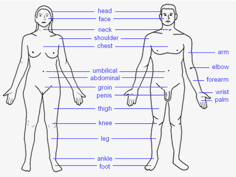 |
HEAD: Forehead – Eye – Ear – Nose – Mouth – Tongue – Teeth – Jaw – Face – Cheek – Chin TORSO: Shoulders – Spine – Chest – Breast – Ribcage – Abdomen – Belly button LIMBS: Arm – Elbow – Forearm – Wrist – Hand – Finger (Thumb - Index finger - Middle finger - Ring finger - Little finger) – Leg – Lap – Thigh – Knee – Calf – Heel – Ankle – Foot – Toe (Hallux) |
Joints and ligaments of upper limbs | |
|---|---|
| Shoulder |
sternoclavicular: anterior sternoclavicular - posterior sternoclavicular - interclavicular - costoclavicular acromioclavicular: acromioclavicular - coracoclavicular (trapezoid, conoid) - coracoacromial - superior transverse scapular - inferior transverse of scapula glenohumeral: coracohumeral - glenohumeral (superior, middle, and inferior) - transverse humeral - glenoid labrum |
| Elbow |
proximal radioulnar, humeroradial, humeroulnar: ulnar collateral - radial collateral - annular - oblique cord |
| Forearm |
distal radioulnar: volar radioulnar - dorsal radioulnar wrist/radiocarpal: palmar radiocarpal - dorsal radiocarpal - ulnar collateral - radial collateral |
| Hand |
intercarpal, midcarpal: pisohamate ligament - pisometacarpal ligament carpometacarpal: dorsal carpometacarpal - palmar carpometacarpal intermetacarpal: deep transverse metacarpal - superficial transverse metacarpal metacarpophalangeal, interphalangeal |
Template:Muscles of upper limb
| This page uses Creative Commons Licensed content from Wikipedia (view authors). |