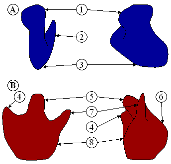
Figure 1: Ribosome structure indicating small subunit (A) and large subunit (B). Side and front view.
(1) Head. (2) Platform. (3) Base. (4) Ridge. (5) Central protuberance. (6) Back. (7) Stalk. (8) Front.
A ribosome is an organelle composed of rRNA and ribosomal proteins (known as a Ribonucleoprotein). It translates mRNA into a polypeptide chain (e.g., a protein). It can be thought of as a factory that builds a protein from a set of genetic instructions. Ribosomes can float freely in the cytoplasm (the internal fluid of the cell) or bind to the endoplasmic reticulum, or to the nuclear envelope. Since ribosomes are ribozymes, it is thought that they might be remnants of the RNA world.
The structure and function of the ribosomes and associated molecules, known as the translational apparatus, has been of research interest since the mid 20th century and is a very active field of study today.
Overview[]
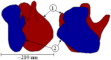
Ribosomes consist of two subunits (Figure 1) that fit together (Figure 2) and work as one to translate the mRNA into a polypeptide chain during protein synthesis (Figure 3). Each subunit consists of one or two very large RNA molecules (known as ribosomal RNA or rRNA) and multiple smaller protein molecules. Crystallographic work has shown that there are no ribosomal proteins close to the reaction site for polypeptide synthesis. This suggests that the protein components of ribosomes act as a scaffold that may enhance the ability of rRNA to synthesise protein rather than directly participating in catalysis.
Figure 2 : Large (1) and small (2) subunit fit together (note this figure mislabels angstroms as nanometers)
Ribosome locations[]
Free ribosomes[]
Free ribosomes occur in all cells, and also in mitochondria and chloroplasts of eukaryotic cells. Free ribosomes usually produce proteins used in the cytosol or organelle in which they occur. As the name implies, they are free in solution and not bound to anything within the cell.
Membrane bound ribosomes[]
When certain proteins are synthesized by a ribosome they can become "membrane-bound". The newly produced polypeptide chains are inserted directly into the ER by the ribosome and are then transported to their destinations. Bound ribosomes usually produce proteins that are used within the cell membrane or are expelled from the cell via exocytosis.
Structure and function[]
The ribosomal subunits of prokaryotes and eukaryotes are quite similar. However, prokaryotes use 70S ribosomes, each consisting of a (small) 30S and a (large) 50S subunit, whereas eukaryotes use 80S ribosomes, each consisting of a (small) 40S and a bound (large) 60S subunit. However, the ribosomes found in chloroplasts and mitochondria of eukaryotes are 70S. [The unit S means Svedberg units, a measure of the rate of sedimentation of a particle in a centrifuge, where the sedimentation rate is associated with the size of the particle. Svedberg units are not additive - two subunits together can have Svedberg values that do not add up to that of the entire ribosome]. Also, the 70S ribosomes are vulnerable to some antibiotics that the 80S ribosomes are not. This helps pharmaceutical companies create drugs that can destroy a bacterial infection without harming the animal/human host's cells.
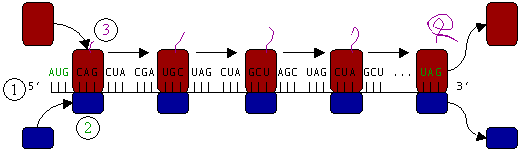
Figure 3 : Translation of mRNA (1) by a ribosome (2) into a polypeptide chain (3). The mRNA begins with a start codon (AUG) and ends with a stop codon (UAG).
In Figure 3, both ribosomal subunits (small and large) assemble at the start codon (the 5' end of the mRNA). The ribosome uses tRNA (transfer RNAs which are RNA molecules that carry an amino acid and present the matching anti-codon, according to the genetic code, to the ribosome) which matches the current codon (triplet) on the mRNA to append an amino acid to the polypeptide chain. This is done for each triplet on the mRNA, while the ribosome moves towards the 3' end of the mRNA. Usually in bacterial cells, several ribosomes are working parallel on a single mRNA. this is what we call polyribosome or polysome.
Atomic structure[]

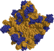
Atomic Structure of the 50S Subunit
The atomic structure of the 50S large subunit ribosome from the archeal, Haloarcula marismortui was published in Science on August 11, 2000 by N. Ban et al.
Soon after the structure of the 30S from Thermus thermophilus was published in Cell on September 1, 2000, by F. Schluenzen et. al.. Shortly after a more detailed structure was published in Nature on September 21, 2000 by B. T. Wimberly, et al..
Using these coordinates, M. M. Yusupov, et al. were able to reconstruct the entire Thermus thermophilus 70S particle at low resolution, which was published in Science in May 2001.
Assessment |
Biopsychology |
Comparative |
Cognitive |
Developmental |
Language |
Individual differences |
Personality |
Philosophy |
Social |
Methods |
Statistics |
Clinical |
Educational |
Industrial |
Professional items |
World psychology |
Biological: Behavioural genetics · Evolutionary psychology · Neuroanatomy · Neurochemistry · Neuroendocrinology · Neuroscience · Psychoneuroimmunology · Physiological Psychology · Psychopharmacology (Index, Outline)
See also[]
Translation
Prokaryotic translation
Eukaryotic translation
External link[]
- 70S Ribosome Architecture Animation of a working Ribosome
| Organelles of the cell |
|---|
| Acrosome | Chloroplast | Cilium/Flagellum | Centriole | Endoplasmic reticulum | Golgi apparatus | Lysosome | Melanosome | Mitochondrion | Myofibril | Nucleus | Parenthesome | Peroxisome | Plastid | Ribosome | Vacuole | Vesicle |
This article contains material from the Science Primer published by the NCBI, which, as a US government publication, is in the public domain at https://www.ncbi.nlm.nih.gov/home/about/policies/.
ca:Ribosoma cs:Ribozom da:Ribosom de:Ribosom es:Ribosoma eo:Ribosomo fa:ریبوزوم fr:Ribosome ko:리보솜 he:ריבוזום la:Ribosoma lt:Ribosoma lb:Ribosom mk:Рибозом nl:Ribosoom no:Ribosom pt:Ribossomo ru:Рибосома sl:Ribosom sr:Рибозом sv:Ribosom vi:Ribosome uk:Рибосома zh:核糖体
| This page uses Creative Commons Licensed content from Wikipedia (view authors). |