{Biopsy}}
| Brain: Rhomboid fossa | ||
|---|---|---|
 | ||
| Hind-brain of a human embryo of three months—viewed from behind and partly from left side. (Rhomboid fossa labeled at center.) | ||
 | ||
| Rhomboid fossa. | ||
| Latin | f. rhomboidea | |
| Gray's | subject #187 798 | |
| Part of | ||
| Components | ||
| Artery | ||
| Vein | ||
| BrainInfo/UW | - | |
| MeSH | [1] | |
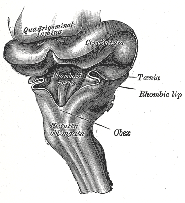
The anterior part of the fourth ventricle is named, from its shape, the rhomboid fossa, and its anterior wall, formed by the back of the pons and medulla oblongata, constitutes the floor of the fourth ventricle.
It is covered by a thin layer of gray substance continuous with that of the medulla spinalis; superficial to this is a thin lamina of neuroglia which constitutes the ependyma of the ventricle and supports a layer of ciliated epithelium.
The fossa consists of three parts, superior, intermediate, and inferior.
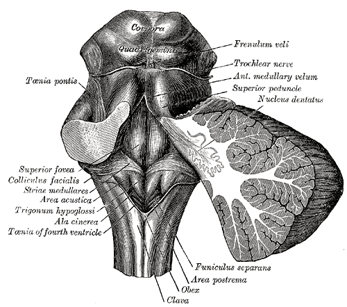
- The superior part is triangular in shape and limited laterally by the superior cerebellar peduncle; its apex, directed upward, is continuous with the cerebral aqueduct; its base is represented by an imaginary line at the level of the upper ends of the superior foveae.
- The intermediate part extends from this level to that of the horizontal portions of the taeniae of the ventricle; it is narrow above where it is limited laterally by the middle peduncle, but widens below and is prolonged into the lateral recesses of the ventricle.
- The inferior part is triangular, and its downwardly directed apex, named the calamus scriptorius, is continuous with the central canal of the closed part of the medulla oblongata.
The sulcus limitans forms the lateral boundary of the medial eminence.
In the superior part of the rhomboid fossa it corresponds with the lateral limit of the fossa and presents a bluish-gray area, the locus cæruleus, which owes its color to an underlying patch of deeply pigmented nerve cells, termed the substantia ferruginea.
At the level of the colliculus facialis the sulcus limitans widens into a flattened depression, the superior fovea, and in the inferior part of the fossa appears as a distinct dimple, the inferior fovea.
Lateral to the foveæ is a rounded elevation named the area acustica, which extends into the lateral recess and there forms a feebly marked swelling, the tuberculum acusticum.
Winding around the inferior peduncle and crossing the area acustica and the medial eminence are a number of white strands, the striæ medullares, which form a portion of the cochlear division of the acoustic nerve and disappear into the median sulcus.
Below the inferior fovea, and between the trigonum hypoglossi and the lower part of the area acustica is a triangular dark field, the ala cinerea, which corresponds to the sensory nucleus of the vagus and glossopharyngeal nerves.
The lower end of the ala cinerea is crossed by a narrow translucent ridge, the funiculus separans, and between this funiculus and the clava, is a small tongue-shaped area, the area postrema.
On section it is seen that the funiculus separans is formed by a strip of thickened ependyma, and the area postrema by loose, highly vascular, neuroglial tissue containing nerve cells of moderate size.
Additional images[]
")
")
This article was originally based on an entry from a public domain edition of Gray's Anatomy. As such, some of the information contained herein may be outdated. Please edit the article if this is the case, and feel free to remove this notice when it is no longer relevant.
| This page uses Creative Commons Licensed content from Wikipedia (view authors). |