Assessment |
Biopsychology |
Comparative |
Cognitive |
Developmental |
Language |
Individual differences |
Personality |
Philosophy |
Social |
Methods |
Statistics |
Clinical |
Educational |
Industrial |
Professional items |
World psychology |
Biological: Behavioural genetics · Evolutionary psychology · Neuroanatomy · Neurochemistry · Neuroendocrinology · Neuroscience · Psychoneuroimmunology · Physiological Psychology · Psychopharmacology (Index, Outline)
| Nerve: Pterygopalatine ganglion | ||
|---|---|---|
 | ||
| Alveolar branches of superior maxillary nerve and pterygopalatine ganglion. (Sphenopalatine ganglion labeled at center top.) | ||
 | ||
| The pterygopalatine ganglion and its branches. (Pterygopalatine ganglion visible but not labeled, as large yellow ganglion in upper-right center.) | ||
| Latin | g. pterygopalatinum | |
| Gray's | subject #200 891 | |
| Innervates | ||
| From | trigeminal nerve | |
| To | ||
| MeSH | [1] | |
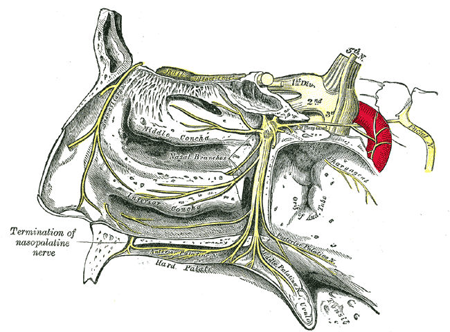
The pterygopalatine ganglion (or sphenopalatine ganglion) is a parasympathetic ganglion found in the pterygopalatine fossa.
Structure[]
The pterygopalatine ganglion (of Meckel), the largest of the parasympathetic ganglia associated with the branches of the trigeminal nerve, is deeply placed in the pterygopalatine fossa, close to the sphenopalatine foramen. It is triangular or heart-shaped, of a reddish-gray color, and is situated just below the maxillary nerve as it crosses the fossa.
The pterygopalatine ganglion supplies the lacrimal gland, paranasal sinuses, glands of the mucosa of the nasal cavity and pharynx, the gingiva, and the mucous membrane and glands of the hard palate. It communicates anteriorly with the nasopalatine nerve.
Roots[]
It receives a sensory, a motor, and a sympathetic root.
Sensory root[]
Its sensory root is derived from two sphenopalatine branches of the maxillary nerve; their fibers, for the most part, pass directly into the palatine nerves; a few, however, enter the ganglion, constituting its sensory root.
Motor root[]
Its motor root is derived from the nervus intermedius (a part of the facial nerve) through the greater petrosal nerve.
In the pterygopalatine ganglion, the parasympathetic fibers they form synapses with neurons whose postganglionic axons, vasodilator and secretory fibers, are distributed with the deep branches of the trigeminal nerve to the mucous membrane of the nose, soft palate, tonsils, uvula, roof of the mouth, upper lip and gums, and to the upper part of the pharynx. It also sends postganglionic parasympathetic fibers to the lacrimal gland via the zygomatic nerve, a branch of the maxillary nerve (from the trigeminal nerve) which then connects with the lacrimal nerve (a branch of the ophthalmic nerve, also part of the trigeminal nerve) to arrive at the lacrimal gland.
Sympathetic root[]
The ganglion also consists of sympathetic efferent (postganglionic) fibers from the superior cervical ganglion. These fibers, from the superior cervical ganglion, travel through the carotid plexus, and then through the deep petrosal nerve. The deep petrosal nerve joins with the greater petrosal nerve to form the nerve of the pterygoid canal, which enters the ganglion.
Additional images[]
")
")
")
")
")
External links[]
- Who Named It synd/2132
- Norman/Georgetown cranialnerves (V, VII)
- Dictionary at eMedicine Pterygopalatine+ganglion
This article was originally based on an entry from a public domain edition of Gray's Anatomy. As such, some of the information contained herein may be outdated. Please edit the article if this is the case, and feel free to remove this notice when it is no longer relevant.
I-IV: olfactory - optic - oculomotor - trochlear
V: trigeminal: trigeminal ganglion
V1: ophthalmic: lacrimal - frontal (supratrochlear, supraorbital) - nasociliary (long root of ciliary, long ciliary, infratrochlear, posterior ethmoidal, anterior ethmoidal) - ciliary ganglion (short ciliary)
V2: maxillary: middle meningeal - in the pterygopalatine fossa (zygomatic, zygomaticotemporal, zygomaticofacial, sphenopalatine, posterior superior alveolar)
in the infraorbital canal/infraorbital nerve (middle superior alveolar, anterior superior alveolar)
on the face (inferior palpebral, external nasal, superior labial, infraorbital plexus) - pterygopalatine ganglion (deep petrosal, nerve of pterygoid canal)
branches of distribution (palatine, nasopalatine, pharyngeal)
V3: mandibular: nervus spinosus - medial pterygoid - anterior (masseteric, deep temporal, buccal, lateral pterygoid)
posterior (auriculotemporal, lingual, inferior alveolar, mylohyoid, mental) - otic ganglion - submandibular ganglion
VI: abducens
VII: facial: nervus intermedius - geniculate - inside facial canal (greater petrosal, nerve to the stapedius, chorda tympani)
at exit from stylomastoid foramen (posterior auricular, digastric - stylohyoid)
on face (temporal, zygomatic, buccal, mandibular, cervical)
VIII: vestibulocochlear: cochlear (striae medullares, lateral lemniscus) - vestibular
IX: glossopharyngeal: fasciculus solitarius - nucleus ambiguus - ganglia (superior, petrous) - tympanic - carotid sinus
X: vagus: ganglia (jugular, nodose) - Alderman's nerve - in the neck (pharyngeal branch, superior laryngeal ext and int, recurrent laryngeal)
in the thorax (pulmonary branches, esophageal plexus) - in the abdomen (gastric plexuses, celiac plexus, gastric plexus)
XI: accessory XII: hypoglossal
Nerves – autonomic nervous system (sympathetic nervous system/ganglion/trunks and parasympathetic nervous system/ganglion) (TA A14.3, GA 9.968) | |||||||
|---|---|---|---|---|---|---|---|
| Head/ cranial |
| ||||||
| Neck/ cervical |
| ||||||
| Chest/ thorax |
| ||||||
| Abdomen/ Lumbar |
| ||||||
| Pelvis/ sacral |
| ||||||
| {| class="navbox collapsible uncollapsed nowraplinks" style="margin:auto; " | |||||||
| |||||||
|}
- de:Ganglion pterygopalatinum
| This page uses Creative Commons Licensed content from Wikipedia (view authors). |