| Brain: Periaqueductal gray | ||
|---|---|---|
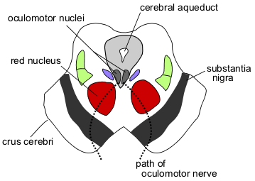 | ||
| Section through superior colliculus showing path of oculomotor nerve. (Periaqueductal gray visible but not labeled.) | ||
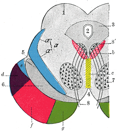 | ||
| Coronal section through mid-brain. 1. Corpora quadrigemina. 2. Cerebral aqueduct. 3. Central gray stratum. 4. Interpeduncular space. 5. Sulcus lateralis. 6. Substantia nigra. 7. Red nucleus of tegmentum. 8. Oculomotor nerve, with 8’, its nucleus of origin. a. Lemniscus (in blue) with a’ the medial lemniscus and a" the lateral lemniscus. b. Medial longitudinal fasciculus. c. Raphé. d. Temporopontine fibers. e. Portion of medial lemniscus, which runs to the lentiform nucleus and insula. f. Cerebrospinal fibers. g. Frontopontine fibers. | ||
| Latin | s. grisea centralis | |
| Gray's | subject #188 806 | |
| Part of | ||
| Components | ||
| Artery | ||
| Vein | ||
| BrainInfo/UW | hier-501 | |
| MeSH | A08.186.211.132.659.822.595 | |
Periaqueductal Gray (PAG; also called the "central gray") is the midbrain grey matter that is located around the cerebral aqueduct within the midbrain. It plays a role in the descending modulation of pain and in defensive behaviour. The ascending pain and temperature fibers of the spinothalamic tract also send information to the PAG via the spinomesencephalic tract. The spinomesencephalic tract is so-named because the fibers orginate in the spine and terminate in the mesencephalon, another name for the midbrain, which is the part of the brain in which the PAG resides.
Role in Analgesia[]
Stimulation of the periaqueductal gray matter of the midbrain activates enkephalin releasing neurons that project to the raphe nuclei in the brainstem. 5-HT (serotonin) released from the raphe nuclei descends to the dorsal horn of the spinal cord where forms excitatory connections with the "inhibitory interneurons" located in Laminae II (aka the substantia gelitanosa). When activated, these interneurons release either enkephalin or dynorphin (endogenous opioid neurotransmitters) which bind to mu opioid receptors* on the axons of incoming C and A-delta fibers carrying pain signals from nociceptors activated in the periphery. The activation of the mu-opioid receptor inhibits the release of substance P from these incoming first order neurons and in turn inhibits the activation of the second order neuron that is responsible for transmitting the pain signal up the spinothalamic tract to the ventroposteriolateral nucleus (VPL) of the thalamus. The nociceptive signal was inhibited before it was able to reach the cortical areas that interpret the signal as "pain" (such as the anterior cingulate). This is sometimes referred to as the "pain gating theory" and is supported by the fact that electrical stimulation of the PAG results in immediate and profound analgesia.
- Three known kinds of opioid receptors have been identified- mu, kappa, and delta. Synthetic opioid and opioid-derivative drugs activate these receptors (possibly by acting on the PAG directly, where a dense amount of these receptors are expressed) to produce analgesia- including heroin, morphine, vicodin, and similar pain modulating compounds.
Role in Defensive Behavior[]
Stimulation of the dorsal and lateral aspects of the PAG [in the rat] can provoke fight or flight response characterised by freezing, running, jumping, tachycardia and increases in blood pressure and muscle tonus. Conversely, stimulation of the caudal ventrolateral PAG can result in an immobile, relaxed posture known as quiescence.
Lesions of the caudal ventrolateral PAG can greatly reduce conditioned freezing, while lesions of the dorsal aspect can reduce innate defensive behavior, virtually "taming" the animal.
Role in Reproductive Behavior[]
Neurons of the PAG are excited by endorphins and by opiate analgesics. It also plays a role in female copulatory behavior (see Lordosis behavior) via a pathway from the ventromedial nucleus of the thalamus.
External links[]
| This page uses Creative Commons Licensed content from Wikipedia (view authors). |