Assessment |
Biopsychology |
Comparative |
Cognitive |
Developmental |
Language |
Individual differences |
Personality |
Philosophy |
Social |
Methods |
Statistics |
Clinical |
Educational |
Industrial |
Professional items |
World psychology |
Biological: Behavioural genetics · Evolutionary psychology · Neuroanatomy · Neurochemistry · Neuroendocrinology · Neuroscience · Psychoneuroimmunology · Physiological Psychology · Psychopharmacology (Index, Outline)
| Brain: Paraventricular nucleus of hypothalamus | ||
|---|---|---|
 | ||
| Human paraventricular nucleus (PVN) in this coronal section is indicated by the shaded area. Dots represent vasopressin (AVP) neurons (also seen in the supraoptic nucleus, SON). The medial surface is the 3rd ventricle (3V), with more lateral to the left. | ||
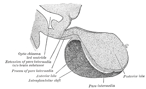 | ||
| Magnocellular neurons of the PVN and SON project to the posterior "lobe" of the pituitary | ||
| Latin | nuclei paraventricularis hypothalami | |
| Gray's | subject # | |
| Part of | ||
| Components | ||
| Artery | ||
| Vein | ||
| BrainInfo/UW | hier-370 | |
| MeSH | A08.186.211.730.385.357.342.400 | |
The paraventricular nucleus (PVN) is an aggregation of neurons in the hypothalamus which produces many hormones.
Location[]
It is adjacent to the third ventricle (hence the name of the nucleus.)
Although it is in the periventricular zone, it is not to be confused with the periventricular nucleus that occupies a more medial, subjacent position to the third ventricle.
The PVN is highly vascularised and is within the blood-brain barrier, although the neuroendocrine neurons in this nucleus project to sites (the median eminence and the posterior pituitary) that lack a blood-brain barrier.
Neurons[]
The PVN contains magnocellular neurosecretory cells whose axons extend into the posterior pituitary, parvocellular neurosecretory cells that project to the median eminence, and several populations of peptide-containing cells that project to many different brain regions.
Magnocellular neurosecretory neurons[]
The magnocellular cells in the PVN produce two hormones:
These peptide hormones are packaged in large dense-core vesicles, which are transported down the axons and released from neurosecretory nerve terminals in the posterior pituitary gland.
Similar magnocellular neurons are found in the supraoptic nucleus.
Parvocellular neurosecretory neurons[]
The parvocellular neurosecretory neurons of the PVN project axons to the median eminence, at the base of the brain. At the median eminence, the neurosecretory nerve terminals release peptides into the blood vessels of the hypothalamo-pituitary portal system. These vessels carry the peptides to the anterior pituitary gland, where they regulate hormone secretion into the systemic circulation. The parvocellular neurosecretory cells include cells which make the following hormones:
- corticotropin-releasing hormone (CRH), which regulates ACTH secretion from the anterior pituitary gland,
- vasopressin: vasopressin released from these neurons also regulates ACTH secretion; vasopressin and CRH act synergistically to stimulate ACTH secretion.
- thyrotropin-releasing hormone (TRH), which regulates TSH secretion.
Centrally-projecting neurons[]
As well as neuroendocrine neurons, the PVN contains interneurons and populations of neurons that project centrally (i.e. to other brain regions). The centrally-projecting neurons include:
- parvocellular oxytocin cells that project mainly to the brainstem and spinal cord. The oxytocin cells that project to the brainstem are involved in gastric reflexes, those that project to the spinal cord are involved in penile erection.
- parvocellular vasopressin cells that project to many areas of the hypothalamus and limbic system, as well as to the brainstem and spinal cord. These neurons are involved in blood pressure regulation and thermoregulation.
- parvocellular CRH neurons that are thought to be involved in stress-associated behaviors
Afferent inputs to the PVN[]
The PVN receives afferent inputs from many brain regions.
Amongst these, inputs from neurons in structures adjacent to the anterior wall of the third ventricle ("AV3V region") carry information about the electrolyte composition of the blood, and about circulating concentrations of hormones such as angiotensin and relaxin to regulate the magnocellular neurons.
Inputs from the brainstem nucleus of the solitary tract and the ventrolateral medulla carry information from the heart and stomach. Inputs from the hippocampus to the CRH neurones are important regulators of stress responses.
Inputs from neuropeptide Y-containing neurons in the arcuate nucleus co-ordinate metabolic regulation via TRH secretion with regulation of energy intake.
References[]
External links[]
| This page uses Creative Commons Licensed content from Wikipedia (view authors). |