| Brain: Orbitofrontal cortex | ||
|---|---|---|
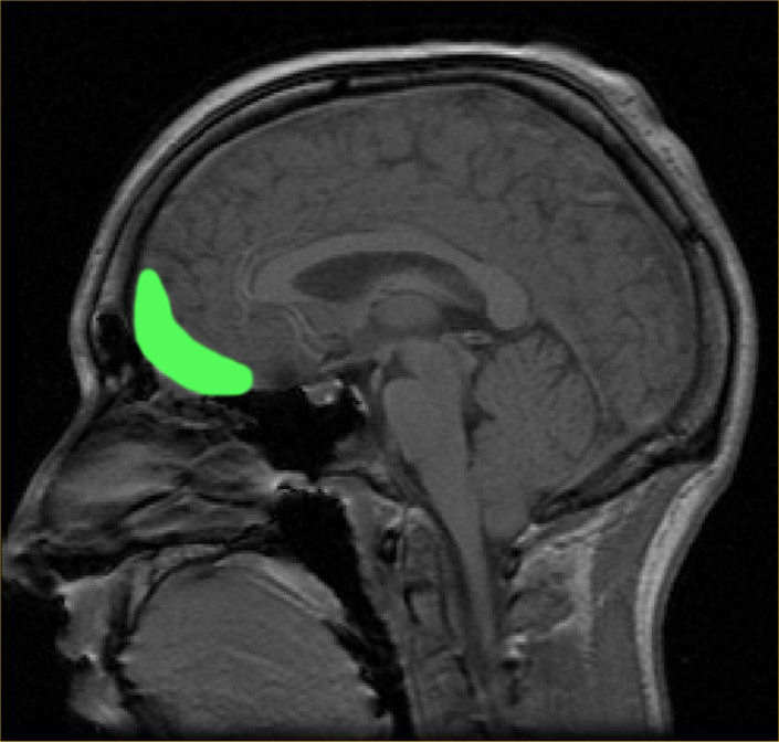 | ||
| Approximate location of the OFC shown on a saggital MRI | ||
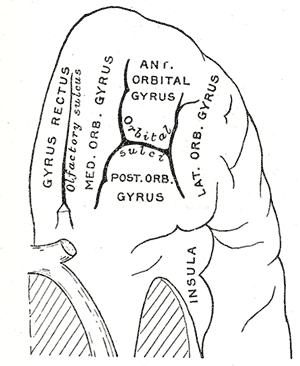 | ||
| Orbital surface of left frontal lobe. | ||
| Latin | ' | |
| Gray's | subject #189 822 | |
| Part of | ||
| Components | ||
| Artery | ||
| Vein | ||
| BrainInfo/UW | hier-73 | |
| MeSH | [1] | |
The orbitofrontal cortex (OFC) is a region of association cortex of the human brain involved in cognitive processes such as decision making. This region is named based upon its location within the frontal lobes, resting above the orbits of the eyes. Because of its functions in emotion, the orbitofrontal cortex is considered to be a part of the limbic system.
Normal functioning of the OFC[]
The OFC is thought to regulate planning behavior associated with sensitivity to reward and punishment. [1]. There are two themes of evidence to support this theory; research in healthy participants, and research in patients with damage to the OFC.
Research methods examining OFC dysfunction[]
- The visual discrimination test[2] has two components: reversal learning and extinction. In "reversal learning", the participant is presented with one of two pictures, A and B. They learn that they will be rewarded if they press a button when picture A is displayed, but punished if they press the button when picture B is displayed. Once this rule has been established, the rule swaps. In other words, now it is correct to press the button for picture B, not picture A. Most healthy participants pick up on this rule reversal almost immediately, but patients with OFC damage continue to respond to the original pattern of reinforcement although they are now being punished for persevering with it. Rolls et al noted that this pattern of behaviour was particularly unusual given that the patients reported that they understood the rule.
- The second component of the test is "extinction". Again, participants learn to press the button for picture A but not picture B. However this time, instead of the rules reversing, the rule changes altogether. Now the participant will be punished for pressing the button in response to either picture. The correct response is not to press the button at all, but people with OFC dysfunction find it difficult to resist the temptation to press the button despite being punished for it.
- The Iowa gambling task is a simulation of real life decision-making[1] which is widely used in research of cognition and emotion. Participants are presented with 4 virtual decks of cards on a computer screen. They are told that each time they choose a card they will win some game money. Every so often, however, when they choose a card they will win money, but will also lose some money too. They are told that the aim of the game is to win as much money as possible. The task is meant to be opaque, i.e. participants are not meant to consciously work out the rule, and they are supposed to choose cards based on their "gut reaction". Two of the decks are "bad decks", which means that, over a long enough time, they will make a net loss; the other two decks are "good decks" and will make a net gain over time. Most healthy participants sample cards from each deck, and after about 40 or 50 selections are fairly good at sticking to the good decks. Patients with OFC dysfunction, however, continue to perseverate with the bad decks, sometimes even though they know that they are losing money overall. Concurrent measurement of galvanic skin response shows that healthy participants show a "stress" reaction to hovering over the bad decks after only 10 trials, long before conscious sensation that the decks are bad. By contrast, patients with OFC dysfunction never develop this physiological reaction to impending punishment. Bechara and his colleagues explain this in terms of the somatic marker hypothesis. The Iowa gambling task is currently being used by a number of research groups (including the Institute of Psychiatry and Institute of Neurology) using fMRI to investigate which brain regions are activated by the task in healthy volunteers as well as clinical groups with conditions such as schizophrenia and obsessive compulsive disorder.
- The Faux pas test is a series of vignettes recounting a social occasion where someone said something they shouldn't have said, or something awkward. The participant's task is to identify what was said that was awkward, why it was awkward, how people would have felt in reaction to the faux pas, and a factual control question. Although first designed for use in people on the autistic spectrum[3], the test is also sensitive to patients with OFC dysfunction, who cannot judge when something socially awkward has happened despite appearing to understand the story perfectly well.
Everyday consequences of damage to the OFC[]
Destruction of the OFC through acquired brain injury typically leads to a pattern of disinhibited behaviour. Examples include swearing excessively, hypersexuality, poor social interaction, compulsive gambling, excessive alcohol / smoking / drug use and poor empathising ability. Disinhibited behaviour by patients with some forms of frontotemporal dementia is thought to be caused by degeneration of the OFC[4]. Patients with damage to the OFC tend to make rash decisions and typically manage their finances poorly.
References[]
- ↑ 1.0 1.1 Bechara A, Damasio AR, Damasio H, Anderson SW. Insensitivity to future consequences following damage to human prefrontal cortex. Cognition 1994; 50: 7-15.
- ↑ Rolls ET, Hornak J, Wade D, McGrath J. Emotion-related learning in patients with social and emotional changes associated with frontal lobe damage. J Neurol Neurosurg Psychiatry 1994; 57: 1518-1524.
- ↑ Stone VE, Baron-Cohen S, Knight RT. Frontal Lobe Contributions to Theory of Mind. Journal of Medical Investigation 1998a; 10: 640-656.
- ↑ Snowden JS, Bathgate D, Varma A, Blackshaw A, Gibbons ZC, Neary D. Distinct behavioural profiles in frontotemporal dementia and semantic dementia. J Neurol Neurosurg Psychiatry 2001; 70: 323-332.
See also[]
External links[]
| Human brain: Limbic system | |
| Amygdala - Cingulate gyrus - Fornicate gyrus - Hippocampus - Hypothalamus - Mammillary body - Nucleus accumbens - Orbitofrontal cortex - Parahippocampal gyrus |
| This page uses Creative Commons Licensed content from Wikipedia (view authors). |