Assessment |
Biopsychology |
Comparative |
Cognitive |
Developmental |
Language |
Individual differences |
Personality |
Philosophy |
Social |
Methods |
Statistics |
Clinical |
Educational |
Industrial |
Professional items |
World psychology |
Biological: Behavioural genetics · Evolutionary psychology · Neuroanatomy · Neurochemistry · Neuroendocrinology · Neuroscience · Psychoneuroimmunology · Physiological Psychology · Psychopharmacology (Index, Outline)
| Brain: Inferior colliculus | ||
|---|---|---|
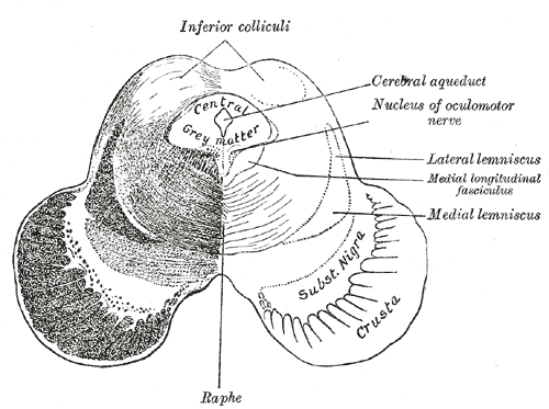 | ||
| Transverse section of mid-brain at level of inferior colliculi. | ||
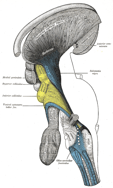 | ||
| Deep dissection of brain-stem. Lateral view. ("Inferior colliculus" is yellow, and is labeled at left. | ||
| Latin | colliculus inferior | |
| Gray's | subject #188 806 | |
| Part of | ||
| Components | ||
| Artery | ||
| Vein | ||
| BrainInfo/UW | hier-467 | |
| MeSH | A08.186.211.132.659.237.364 | |
The inferior colliculi (Latin, lower hill) together with the superior colliculi form the eminences of the corpora quadrigemina, and also part of the tectal region of the midbrain. The inferior colliculus lies caudal to its counterpart - the superior colliculus - above the trochlear nerve, and at the base of the projection of the medial geniculate nucleus (MGN) and the lateral geniculate nucleus (LGN).
The inferior colliculi together with the superior colliculi form the eminences of the corpora quadrigemina, and also part of the tectal region of the midbrain. The inferior colliculus lies caudal to its counterpart - the superior colliculus - above the trochlear nerve, and at the base of the projection of the medial geniculate nucleus (MGN) and the lateral geniculate nucleus (LGN).
Relationship to auditory system[]
The inferior colliculi of the midbrain are located just below the visual processing centers known as the superior colliculi. The inferior colliculus is the first place where vertically orienting data from the fusiform cells in the dorsal cochlear nucleus can finally synapse with horizontally orienting data. This homecoming of the aural dimensions puts these dual mesencephalic bumps in the position to fully integrate all the sound location data.
IC are large auditory nuclei on the right and left sides of the midbrain. It is divided into three parts, the Central Nucleus of IC (CNIC), dorsal cortex and lateral cortex; however, CNIC is the principal way station for ascending auditory information in the IC.
Input and output connections of IC[]
The input connections to the inferior colliculus are composed of many brainstem nuclei. All nuclei except the contralateral ventral nucleus of the lateral lemniscus (LL) send projections to the central nucleus (CNIC) bilaterally. It has been shown that great majority of auditory fibers ascending in the lateral lemniscus terminate in the CNIC. In addition, the IC receives inputs from the auditory cortex, the medial division of the medial geniculate body (MGBm), the posterior limitans (PL), suprapeduncular nucleus (SpN) and subparafascicular (Spf) intralaminar nuclei of the thalamus, the substantia nigra, pars compacta lateralis (SNL), the dorsolateral periaqueductal gray (dlPAG), the nucleus of the brachium of the inferior colliculus (BICN) and deep layers of the superior colliculus (SC).
The inferior colliculus receives input from both the ipsilateral and contralateral cochlear nucleus and respectively the corresponding ears. There is some lateralization, the dorsal projections (containing vertical data) only project to the contralateral inferior colliculus. This inferior colliculus contralateral to the ear it is receiving the most information from, then projects to its ipsilateral medial geniculate nucleus.
The medial geniculate body (MGB) is the output connection from inferior colliculus and the last subcortical way station. The MGB is composed of ventral, dorsal, and medial divisions, which are relatively similar in humans and other mammals. The ventral division receives auditory signals from the central nucleus of the IC.[1]
Function of IC[]
The majority of the ascending fibers from LL project to IC, which means major ascending auditory pathway converge here. IC appears as an integrative station and switchboard as well. It is involved in the integration and routing of multi-modal sensory perception, mainly the startle response and vestibulo-ocular reflex. It is also responsive to specific amplitude modulation frequencies and this might be responsible for detection of pitch. In addition, spatial localization by binaural hearing is a related function of IC as well.
The inferior colliculus has a relatively high metabolism in the brain. The Conrad Simon Memorial Research Initiative measured the blood flow of the IC and put a number at 1.80 cc/g/min in the cat brain. For reference, the runner up in the included measurements was the somatosensory cortex at 1.53. This indicates that the inferior colliculus is metabolically more active than many other parts of the brain. The hippocampus, normally considered to use up a disproportionate amount of energy, was not measured or compared.[2]
Skottun et al. measured the interaural time difference sensitivity of single neurons in the inferior colliculus, and used these to predict behavioural performance. The predicted just noticeable difference was comparable to that achieved by humans in behavioral tests.[3] This suggested that by the level of the inferior colliculus, integration of information over multiple neurons is unnecessary (see population code).
Abbreviations[]
CNIC: Central Nucleus of IC; AVCN: Anterior Ventral Cochlear Nucleus; PVCN: Posterior Ventral Cochlear Nucleus; SCN: Superior Colliculus Nucleus; LSO: Lateral Superior Olive; MSO: Medial Superior Olive; DNLL: Dorsal Nucleus of the Lateral Lemniscus; MGB: Medial Geniculate Body; SC: Superior Colliculus; TB: Trapezoid Body; MGBm: medial division of the medial geniculate body; PL: the posterior limitans nucleus; SpN: suprapeduncular nucleus; Spf: subparafascicular nucleus; SNL: the substantia nigra, pars compacta lateralis; dlPAG: the dorsolateral periaqueductal gray; BICN: the nucleus of the brachium of the inferior colliculus.
See also[]
References[]
- ↑ Gelfand, Stanley A.: Hearing, an Introduction to Psychological and Physiological Acoustics, 4th Ed., Marcel Dekker, 2004, pp. 71-75.
- ↑ Conrad Simon Memorial Research Initiative homepage <http://www.conradsimon.org/InferiorColliculus.shtml>
- ↑ Skottun, Bernt C. et al.: The ability of inferior colliculus neurons to signal differences in interaural delay. PNAS November 20, 2001 vol. 98, no. 24, pp. 14050-14054.
Additional images[]
")
")
")
")
External links[]
Auditory and vestibular pathways | ||||
|---|---|---|---|---|
| Auditory |
inner ear: Hair cells → Spiral ganglion → Cochlear nerve VIII → pons: Cochlear nuclei (Anterior, Dorsal) → Trapezoid body → Superior olivary nuclei → midbrain: Lateral lemniscus → Inferior colliculi → | |||
| Vestibular |
inner ear: Vestibular nerve VIII → pons: Vestibular nuclei (Medial vestibular nucleus, Lateral vestibular nucleus) cerebellum: Flocculonodular lobe spinal cord: Vestibulospinal tract (Medial vestibulospinal tract, Lateral vestibulospinal tract) | |||
| {| class="navbox collapsible nowraplinks" style="margin:auto; " | ||||
| ||||
|} Catgory:Mesencephalon
| This page uses Creative Commons Licensed content from Wikipedia (view authors). |