Assessment |
Biopsychology |
Comparative |
Cognitive |
Developmental |
Language |
Individual differences |
Personality |
Philosophy |
Social |
Methods |
Statistics |
Clinical |
Educational |
Industrial |
Professional items |
World psychology |
Biological: Behavioural genetics · Evolutionary psychology · Neuroanatomy · Neurochemistry · Neuroendocrinology · Neuroscience · Psychoneuroimmunology · Physiological Psychology · Psychopharmacology (Index, Outline)
| Human skull | ||
|---|---|---|
| Human skulls side simplified | ||
| Latin | cranium | |
| Gray's | subject # | |
| System | ||
| MeSH | [1] | |
| Human skull front bones | ||
.svg)
In humans, the adult skull is normally made up of 22 bones.[1] Except for the mandible, all of the bones of the skull are joined together by sutures, synarthrodial (immovable) joints formed by bony ossification, with Sharpey's fibres permitting some flexibility.
Components[]
The lower inner surface of the neurocranium
Eight bones form the neurocranium (brain case), a protective vault of bone surrounding the brain and brain stem. Fourteen bones form the splanchnocranium, which comprises the bones supporting the face. Encased within the temporal bones are the six auditory ossicles of the middle ear. The hyoid bone, supporting the larynx, is usually not considered as part of the skull, as it is the only bone that does not articulate with other bones of the skull.
The skull also contains the sinus cavities, which are air-filled cavities lined with respiratory epithelium, which also lines the large airways. The exact functions of the sinuses are debatable; they contribute to lessening the weight of the skull with a minimal reduction in strength, they contribute to resonance of the voice, and assist in the warming and moistening of air drawn in through the nasal cavity.
Development of the skull[]
The lower inner surface of the neurocranium- 11 weeks' fertilization age
The skull is a complex structure; its bones are formed both by intramembranous and endochondral ossification. The skull roof, comprising the bones of the splanchnocranium (face) and the sides and roof of the neurocranium, are formed by intramembranous (or dermal) ossification, though the temporal bones are formed by endochondral ossification. The endocranium, the bones supporting the brain (the occipital, sphenoid, and ethmoid) are largely formed by endochondral ossification. Thus frontal and parietal bones are purely membranous.[2] The geometry of the cranial base and its fossas: anterior, middle and posterior changes rapidly, especially during the first trimester of pregnancy. The first trimester is crucial for development of skull defects.[3]
At birth, the human skull is made up of 404 separate bony elements. As growth occurs, many of these bony elements gradually fuse together into solid bone (for example, the frontal bone). The bones of the roof of the skull are initially separated by regions of dense connective tissue called "fontanels". There are six fontanels: one anterior (or frontal), one posterior (or occipital), two sphenoid (or anterolateral), and two mastoid (or posterolateral). At birth these regions are fibrous and moveable, necessary for birth and later growth. This growth can put a large amount of tension on the "obstetrical hinge", which is where the squamous and lateral parts of the occipital bone meet. A possible complication of this tension is rupture of the great cerebral vein of Galen. As growth and ossification progress, the connective tissue of the fontanelles is invaded and replaced by bone creating sutures. The five sutures are the two squamous, one coronal, one lambdoid, and one sagittal sutures. The posterior fontanel usually closes by eight weeks, but the anterior fontanel can remain open up to eighteen months. The anterior fontanel is located at the junction of the frontal and parietal bones; it is a "soft spot" on a baby's forehead. Careful observation will show that you can count a baby's heart rate by observing his or her pulse pulsing softly through the anterior fontanel.
Pathology[]
If the brain is bruised or injured it can be life-threatening. Normally the skull protects the brain from damage through its hard unyieldingness; the skull is one of the most durable substances found in nature with it needing the force of about 1 ton to reduce the diameter of the skull by 1 cm.[4] In some cases, however, of head injury, there can be raised intracranial pressure through mechanisms such as a subdural haematoma. In these cases the raised intracranial pressure can cause herniation of the brain out of the foramen magnum ("coning") because there is no space for the brain to expand; this can result in significant brain damage or death unless an urgent operation is performed to relieve the pressure. This is why patients with concussion must be watched extremely carefully.
Dating back to Neolithic times, a skull operation called trepanation was sometimes performed. This involved drilling holes in the cranium. Examination of skulls from this period reveals that the "patients" sometimes survived for many years afterward. It seems likely that trepanation was performed for ritualistic or religious reasons and not only as an attempted life-saving technique.
Craniometry and morphology of human skulls[]
Like the face of a living individual, a human skull and teeth can also tell, to a certain degree, the life history and origin of its owner. Forensic scientists and archaeologists use metric and nonmetric traits to estimate what the bearer of the skull looked like. When a significant amount of bones are found, such as at Spitalfields in the UK and Jōmon shell mounds in Japan, osteologists can use traits, such as the proportions of length, height and width, to know the relationships of the population of the study with other living or extinct populations.
The German physician Franz Joseph Gall in around 1800 formulated the theory of phrenology, which attempted to show that specific features of the skull are associated with certain personality traits or intellectual capabilities of its owner. This theory is now considered to be obsolete.
Sexual dimorphism[]
In general, male skulls tend to be larger and more robust than female skulls, which are more gracile.[citation needed] Male skulls typically have more prominent supraorbital ridges, a more prominent glabella, and more prominent temporal lines. Male skulls on average have larger, broader palates, squarer orbits, larger mastoid processes, larger sinuses, and larger occipital condyles than those of females. Male mandibles typically have squarer chins and thicker, rougher muscle attachments than female mandibles.
Partial human skulls
All of these features vary considerably within human populations, making it difficult to identify the sex of a skull without knowledge of the population from which it came.
Ancestry[]
Among archaeologists and forensic scientists, it is stated that the most consistent and unique trait of ancestry in a skeleton is its skull shape (see craniometry).[citation needed]
Additional images[]
|
| Human skulls
]]See also[]
- Skull, a general article on skulls other than the human skull
- Base of the skull, detailed list of the anatomical structures found at the base of the skull
- Craniometry
- Foramina of the skull, list of holes (foramina) in the base of the skull
- Head and neck anatomy
- Phrenology, the pseudoscientific process of determining personality from the shape of the head
- Plagiocephaly, the abnormal flattening of one side of the skull
- Human skull symbolism
- Anatomical terms of location
References[]
- ↑ Skull Basics
- ↑ Carlson, Bruce M. (1999). Human Embryology & Developmental Biology, 166–170, Mosby.
- ↑ Derkowski W; Kedzia A; Glonek M. Clinical anatomy of the human anterior cranial fossa during the prenatal period. Folia morphologica 2003;62(3):271-3,PMID: 14507064.
- ↑ Holbourn, A. H. S. (1943). MECHANICS OF HEAD INJURIES. The Lancet, 242: (6267), 438-441. DOI:10.1016/S0140-6736(00)87453-X
Human cranial bones
|
|---|
|
occipital bone: Foramen magnum | Squama occipitalis (Inion | Nuchal lines | Planum occipitale | Planum nuchale | Cruciform eminence | Internal occipital protuberance | Sagittal sulcus | Internal occipital crest) parietal bone: Parietal eminence | Temporal line | Parietal foramen frontal bone: Squama frontalis (Frontal suture | Frontal eminence | Superciliary arches | Glabella | Supraorbital foramen | Zygomatic process | Sagittal sulcus | Frontal crest | Foramen cecum) temporal bone: Squama temporalis (Articular tubercle | Suprameatal triangle | Mandibular fossa | Petrotympanic fissure) | Mastoid portion (Mastoid foramen sphenoid bone: Sphenoidal sinuses | Ethmoidal spine | Optic foramen | Sella turcica | Fossa hypophyseos | Dorsum sellae | Posterior clinoid processes | Carotid groove | Lingula sphenoidalis | Sphenoidal conchæ | Great wings (Spina angularis | Foramen rotundum | Foramen ovale | Foramen Vesalii | Foramen spinosum | Infratemporal crest | Sulcus tubae auditivae | Small wings | Superior orbital fissure | Anterior clinoid process | Optic foramen) ethmoid bone: Cribriform plate | Crista galli | Perpendicular plate | Labyrinth | Ethmoid sinus | Uncinate process | Middle nasal concha | Superior meatus | Superior nasal concha | Middle meatus |
Bones of head and neck: the face | |
|---|---|
| maxilla |
Body of maxilla - Maxillary sinus - surfaces of body Anterior (Incisive fossa, Canine fossa, Infraorbital foramen, Anterior nasal spine) - Infratemporal (Alveolar canals, Maxillary tuberosity) - Orbital (Infraorbital groove, Infraorbital canal) - Nasal (Pterygopalatine canal) |
| lacrimal |
Posterior lacrimal crest - Lacrimal groove |
| zygomatic |
Orbital process - foramina (Zygomaticofacial, Zygomaticotemporal, Zygomaticoörbital) |
| palatine |
Pterygopalatine fossa - Pterygoid fossa |
| mandible |
body (Symphysis menti, Mental protuberance, Mental foramen, Mental spine, Mylohyoid line) |
| others |
nasal bone - inferior nasal conchae (ethmoidal process, maxillary process) - vomer |
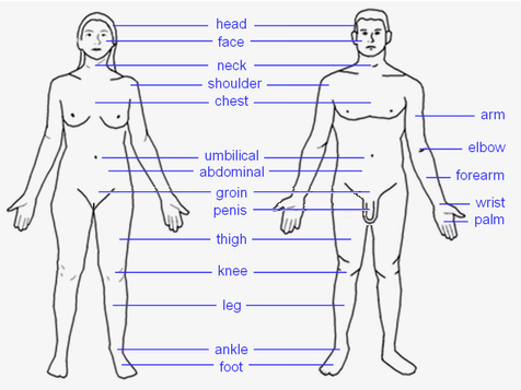 |
HEAD: Forehead – Eye – Ear – Nose – Mouth – Tongue – Teeth – Jaw – Face – Cheek – Chin TORSO: Shoulders – Spine – Chest – Breast – Ribcage – Abdomen – Belly button LIMBS: Arm – Elbow – Forearm – Wrist – Hand – Finger (Thumb - Index finger - Middle finger - Ring finger - Little finger) – Leg – Lap – Thigh – Knee – Calf – Heel – Ankle – Foot – Toe (Hallux) |
| This page uses Creative Commons Licensed content from Wikipedia (view authors). |