No edit summary |
No edit summary |
||
| (2 intermediate revisions by 2 users not shown) | |||
| Line 1: | Line 1: | ||
{{BioPsy}} |
{{BioPsy}} |
||
| + | {{Infobox Brain| |
||
| ⚫ | |||
| + | Name = Rhombencephalon | |
||
| + | GraySubject = 187 | |
||
| + | GrayPage = 767 | |
||
| + | Image = EmbryonicBrain.svg | |
||
| + | Caption = Diagram depicting the main subdivisions of the embryonic vertebrate brain. These regions will later differentiate into [[forebrain]], [[midbrain]] and [[hindbrain]] structures. | |
||
| + | Image2 = Gray682.png | |
||
| ⚫ | |||
| + | IsPartOf = | |
||
| + | Components = | |
||
| + | Artery = | |
||
| + | Vein = | |
||
| + | BrainInfoType = hier | |
||
| + | BrainInfoNumber = 531 | |
||
| + | MeshName = Rhombencephalon | |
||
| + | MeshNumber = A08.186.211.132.810 | |
||
| + | DorlandsPre = r_12 | |
||
| + | DorlandsSuf = 12709581 | |
||
| + | }} |
||
The '''rhombencephalon''' (or '''hindbrain''') is a [[Morphogenesis|developmental]] categorization of portions of the [[central nervous system]] in [[vertebrates]]. |
The '''rhombencephalon''' (or '''hindbrain''') is a [[Morphogenesis|developmental]] categorization of portions of the [[central nervous system]] in [[vertebrates]]. |
||
| − | The rhombencephalon can be subdivided in a variable number of transversal swellings called |
+ | The rhombencephalon can be subdivided in a variable number of transversal swellings called [[rhombomere]]s. In the human embryo we can distinguish eight rhombomeres, from caudal to rostral: Rh7-Rh1 and the [[List of anatomical isthmi| isthmus]] (the most [[Anatomical_terms_of_location|rostral]] rhombomere). |
| + | A rare disease of the rhombencephalon, "rhombencephalosynapsis" is characterized by a missing [[vermis]] resulting in a fused cerebellum. Patients generally present with [[cerebellar ataxia]]. |
||
| − | The myelencephalon forms the [[medulla]] in the adult brain; it contains a portion of the [[fourth ventricle]], the [[glossopharyngeal nerve]] (CN IX), [[vagus nerve]] (CN X), [[accessory nerve]] (CN XI), [[hypoglossal nerve]] (CN XII), and a portion of the [[vestibulocochlear nerve]] (CN VIII). |
||
| + | |||
| + | The caudal rhombencephalon has been generally considered as the initiation site for [[neural tube]] closure.<ref>[http://www.springerlink.com/content/f3fc056wde57c1yc/ SpringerLink - Journal Article<!-- Bot generated title -->]</ref> |
||
| + | |||
| + | ==Myelencephalon== |
||
| + | Rhombomeres Rh7-Rh4 form the [[myelencephalon]]. |
||
| + | |||
| + | The myelencephalon forms the [[medulla oblongata]] in the adult brain; it contains: |
||
| + | * a portion of the [[fourth ventricle]], |
||
| + | * the [[glossopharyngeal nerve]] (CN IX), |
||
| + | * [[vagus nerve]] (CN X), |
||
| + | * [[accessory nerve]] (CN XI), |
||
| + | * [[hypoglossal nerve]] (CN XII), |
||
| + | * and a portion of the [[vestibulocochlear nerve]] (CN VIII). |
||
| + | |||
| + | ==Metencephalon == |
||
| + | Rhombomeres Rh3-Rh1 form the [[metencephalon]]. |
||
| + | |||
| + | The metencephalon is composed of the [[pons]] and the [[cerebellum]]; it contains: |
||
| + | * a portion of the fourth ventricle, |
||
| + | * the [[trigeminal nerve]] (CN V), |
||
| + | * [[abducens nerve]] (CN VI), |
||
| + | * [[facial nerve]] (CN VII), |
||
| + | * and a portion of the [[vestibulocochlear nerve]] (CN VIII). |
||
| + | |||
| + | ==Additional images== |
||
| + | <gallery> |
||
| + | Image:Gray18.png|Chick embryo of thirty-three hours’ incubation, viewed from the dorsal aspect. X 30. |
||
| + | Image:Gray40.png|Embryo between eighteen and twenty-one days. |
||
| + | </gallery> |
||
| + | |||
| + | |||
| + | ==See also== |
||
| + | * [[Brain stem]] |
||
| + | * [[Raphe nuclei]] |
||
| + | |||
| + | == References == |
||
| + | <references/> |
||
| − | The metencephalon is composed of the [[pons]] and the [[cerebellum]]; it contains a portion of the fourth ventricle, the [[trigeminal nerve]] (CN V), [[abducens nerve]] (CN VI), [[facial nerve]] (CN VII), and a portion of the [[vestibulocochlear nerve]] (CN VIII). |
||
| − | {{Neuroscience-stub}} |
||
{{Rhombencephalon}} |
{{Rhombencephalon}} |
||
| + | {{Cerebellum}} |
||
| − | [[category:brain]] |
||
| + | |||
| ⚫ | |||
| + | [[Category:Brain]] |
||
[[Category:Brainstem]] |
[[Category:Brainstem]] |
||
[[Category:Central nervous system]] |
[[Category:Central nervous system]] |
||
[[Category:Cranial nerves]] |
[[Category:Cranial nerves]] |
||
[[Category:Developmental biology]] |
[[Category:Developmental biology]] |
||
| + | [[Category:Neuroanatomy]] |
||
| ⚫ | |||
| + | [[Category:Rhombencephalon|*]] |
||
| + | |||
| + | <!-- |
||
[[de:Rhombencephalon]] |
[[de:Rhombencephalon]] |
||
| + | [[es:Rombencéfalo]] |
||
| ⚫ | |||
| + | [[it:Rombencefalo]] |
||
| + | [[nl:Rhombencephalon]] |
||
| ⚫ | |||
| + | --> |
||
| − | {{enWP| Rhombencephalon |
+ | {{enWP| Rhombencephalon}} |
Latest revision as of 07:40, 29 November 2008
Assessment |
Biopsychology |
Comparative |
Cognitive |
Developmental |
Language |
Individual differences |
Personality |
Philosophy |
Social |
Methods |
Statistics |
Clinical |
Educational |
Industrial |
Professional items |
World psychology |
Biological: Behavioural genetics · Evolutionary psychology · Neuroanatomy · Neurochemistry · Neuroendocrinology · Neuroscience · Psychoneuroimmunology · Physiological Psychology · Psychopharmacology (Index, Outline)
| Brain: Rhombencephalon | ||
|---|---|---|
| Diagram depicting the main subdivisions of the embryonic vertebrate brain. These regions will later differentiate into forebrain, midbrain and hindbrain structures. | ||
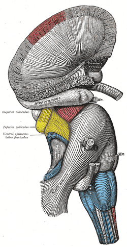
| Superficial dissection of brain-stem. Lateral view. | ||
| Latin | {{{Latin}}} | |
| Gray's | subject #187 767 | |
| Part of | ||
| Components | ||
| Artery | ||
| Vein | ||
| BrainInfo/UW | hier-531 | |
| MeSH | A08.186.211.132.810 | |
The rhombencephalon (or hindbrain) is a developmental categorization of portions of the central nervous system in vertebrates.
The rhombencephalon can be subdivided in a variable number of transversal swellings called rhombomeres. In the human embryo we can distinguish eight rhombomeres, from caudal to rostral: Rh7-Rh1 and the isthmus (the most rostral rhombomere).
A rare disease of the rhombencephalon, "rhombencephalosynapsis" is characterized by a missing vermis resulting in a fused cerebellum. Patients generally present with cerebellar ataxia.
The caudal rhombencephalon has been generally considered as the initiation site for neural tube closure.[1]
Myelencephalon
Rhombomeres Rh7-Rh4 form the myelencephalon.
The myelencephalon forms the medulla oblongata in the adult brain; it contains:
- a portion of the fourth ventricle,
- the glossopharyngeal nerve (CN IX),
- vagus nerve (CN X),
- accessory nerve (CN XI),
- hypoglossal nerve (CN XII),
- and a portion of the vestibulocochlear nerve (CN VIII).
Metencephalon
Rhombomeres Rh3-Rh1 form the metencephalon.
The metencephalon is composed of the pons and the cerebellum; it contains:
- a portion of the fourth ventricle,
- the trigeminal nerve (CN V),
- abducens nerve (CN VI),
- facial nerve (CN VII),
- and a portion of the vestibulocochlear nerve (CN VIII).
Additional images
See also
References
Human brain, rhombencephalon, metencephalon: cerebellum (TA 14.1.07, GA 9.788) | |||||||
|---|---|---|---|---|---|---|---|
| Surface anatomy |
| ||||||
| Grey matter |
| ||||||
| White matter |
| ||||||
| This page uses Creative Commons Licensed content from Wikipedia (view authors). |

