Assessment |
Biopsychology |
Comparative |
Cognitive |
Developmental |
Language |
Individual differences |
Personality |
Philosophy |
Social |
Methods |
Statistics |
Clinical |
Educational |
Industrial |
Professional items |
World psychology |
Animals · Animal ethology · Comparative psychology · Animal models · Outline · Index
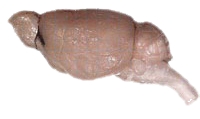
A mouse brain.
Three groups of animals, with some exceptions, have notably complex brains: the arthropods (insects and crustaceans), the cephalopods (octopuses, squid, and similar mollusks), and the craniates (vertebrates)[1]. The brain of arthropods and cephalopods arises from twin parallel nerve cords that extend through the body of the animal. In arthropods, the brain consists of a central brain with three divisions and large optical lobes behind each eye for visual processing[1].
The brain of craniates develops from the anterior section of a single dorsal nerve cord, which later becomes the spinal cord[2]. In craniates, the brain is protected by the bones of the skull. In vertebrates, increasing complexity in the cerebral cortex correlates with height on the phylogenetic and evolutionary tree. Primitive vertebrates such as fish, reptiles, and amphibians have fewer than six layers of neurons in the outer layer of their brains. This cortical configuration is called the allocortex (or heterotypic cortex)[3].
More complex vertebrates such as mammals have developed a six-layered neocortex (or homotypic cortex, neopallium), in addition to having some parts of the brain that are allocortex[3]. In mammals, increasing convolutions of the brain are characteristic of animals with more advanced brains. These convolutions evolved to provide a larger surface area for a greater number of neurons while keeping the volume of the brain compact enough to fit inside the skull. This folding allows for more grey matter to fit into a smaller volume, similar to a really long slinky being able to fit into a tiny box when completed pushed together. The folds of the brain are called gyri, while the spaces between the folds are called the sulci.
Although the general histology of the brain is common to all those who have one, the structural anatomy differs from person to person. Apart from the gross embryological divisions of the brain, the individual location of specific gyri and sulci, primary sensory regions, and other structures in relation to one another differs across creatures.
Invertebrates[]
In insects, the brain can be divided into four parts, the optical lobes, the protocerebrum, the deutocerebrum, and the tritocerebrum. The optical lobes are positioned behind each eye and process visual stimuli [1]. The protocerebrum contains the mushroom bodies, which respond to smell, and the central body complex. In some species such as bees, the mushroom body receives input from the visual pathway as well. The deutocerebrum includes the antennal lobes, which are similar to the mammalian olfactory bulb, and the mechanosensory neuropils which receive information from touch receptors on the head and antennae. The antennal lobes of flies and moths are quite complex.
In cephalopods, the brain is divided into two regions: the supraesophageal mass and the subesophageal mass [1]. These parts are divided by the animal's esophagus. The supra- and subesophageal masses are connected to each other on either side of the esophagus by the basal lobes and the dorsal magnocellular lobes [1]. The large optic lobes are sometimes not considered to be part of the brain proper since the optic lobes are anatomically separate from the brain and are joined to the brain by the optic stalks. However, the optic lobes perform much of the visual processing and functionally can be considered part of the brain.
Vertebrates[]
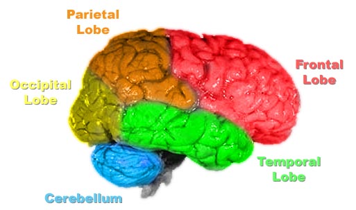
The lobes of the cerebral cortex include the frontal (red), temporal (green), occipital (yellow), and parietal lobes (orange). The cerebellum (blue) is not part of the telencephalon.
In vertebrates a gross division into three major parts is used (see basic list of brain regions below).
The telencephalon (cerebrum) makes up the largest section of the mammalian brain. This is the structure that is most easily visible, and is what most people associate with the “brain”. In humans the fissures (sulci) and convolutions (gyri) give the brain a wrinkled appearance. In most vertebrates the metencephalon is the highest center in the brain, whereas in mammals this role has been adopted by the cerebrum. Because humans walk upright, this creates a flexure, or bent nature, to the brain between the brain stem and the cerebrum that is not present in most other vertebrates. For this reason, descriptions of the locations of certain brain structures in humans as compared to other vertebrate species may be confusing.
Behind (or in humans, below) the cerebrum is the cerebellum. The cerebellum functions mainly in the control of movement and movement timing [2]. It is connected via thick white matter fibers (cerebellar peduncles) to the pons[3]. The cerebrum and the cerebellum each consist of two hemispheres. The telencephalic hemispheres are connected by the corpus callosum, another large white matter tract. An outgrowth of the telencephalon called the olfactory bulb is a major structure in many animals, but in humans and other primates it is relatively small.
Vertebrate nervous systems are distinguished by encephalization and bilateral symmetry. Encephalization refers to the tendency for more complex organisms to gain larger brains through evolutionary time. Larger vertebrates develop a complex system of layered and interconnected neuronal circuitry. In modern species most closely related to the first vertebrates, brains are covered with gray matter that has a three-layer structure (allocortex). Their brains also contain deep brain nuclei and fiber tracts forming the white matter. Most regions of the human cerebral cortex have six layers of neurons (neocortex) [3].
Vertebrate brain regions[]
(See related article at List of regions in the human brain)
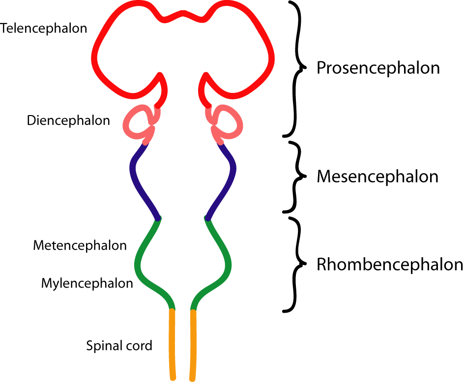
Diagram depicting the main subdivisions of the embryonic vertebrate brain. These regions will later differentiate into forebrain, midbrain and hindbrain structures.
According to the hierarchy based on embryonic and evolutionary development, chordate brains are composed of the three regions that later develop into five total divisions:
- Rhombencephalon (hindbrain)
- Mesencephalon (midbrain)
- Prosencephalon (forebrain)
The brain can also be classified according to function, including divisions such as:
- ↑ 1.0 1.1 1.2 1.3 1.4 Butler, Ann B. (2000). Chordate Evolution and the Origin of Craniates: An Old Brain in a New Head, The Anatomical Record. 261:111-125.
- ↑ 2.0 2.1 Cite error: Invalid
<ref>tag; no text was provided for refs namedkandel - ↑ 3.0 3.1 3.2 3.3 Martin, John H. (1996). Neuroanatomy: Text and Atlas, Second Edition, McGraw-Hill, New York. ISBN 007138183X.