Assessment |
Biopsychology |
Comparative |
Cognitive |
Developmental |
Language |
Individual differences |
Personality |
Philosophy |
Social |
Methods |
Statistics |
Clinical |
Educational |
Industrial |
Professional items |
World psychology |
Biological: Behavioural genetics · Evolutionary psychology · Neuroanatomy · Neurochemistry · Neuroendocrinology · Neuroscience · Psychoneuroimmunology · Physiological Psychology · Psychopharmacology (Index, Outline)
| Nerve: Lateral cutaneous nerve of thigh | ||
|---|---|---|
 | ||
| Plan of lumbar plexus. (Lateral femoral cutaneous visible at left.) | ||
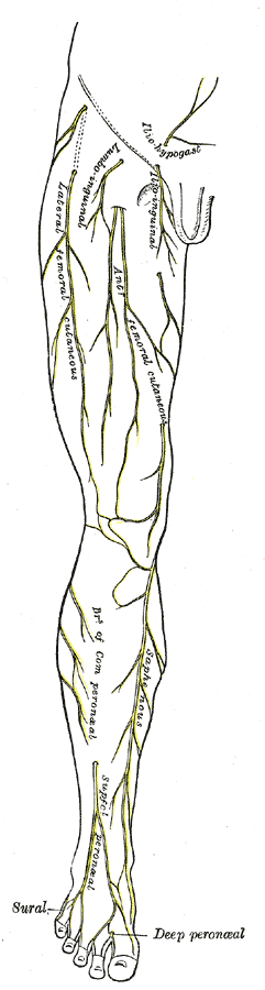 | ||
| Cutaneous nerves of right lower extremity. Front view. | ||
| Latin | nervus cutaneus femoralis lateralis | |
| Gray's | subject #212 953 | |
| Innervates | ||
| From | lumbar plexus (L2–L3) | |
| To | ||
| MeSH | {{{MeshNumber}}} | |
The lateral cutaneous nerve of the thigh (also called the lateral femoral cutaneous nerve) is a cutaneous nerve that innervates the skin on the lateral part of the thigh.
Structure[]
The lateral cutaneous nerve of the thigh is a nerve of the lumbar plexus. It arises from the dorsal divisions of the second and third lumbar nerves. It emerges from the lateral border of the psoas major at about its middle, and crosses the iliacus muscle obliquely, toward the anterior superior iliac spine. It then passes under the inguinal ligament and over the sartorius muscle into the thigh, where it divides into an anterior and a posterior branch.
The anterior branch becomes superficial about 10 cm below the inguinal ligament, and divides into branches which are distributed to the skin of the anterior and lateral parts of the thigh, as far as the knee. The terminal filaments of this nerve frequently communicate with the anterior cutaneous branches of the femoral nerve, and with the infrapatellar branch of the saphenous nerve, forming with them the patellar plexus.
The posterior branch pierces the fascia lata, and subdivides into filaments which pass backward across the lateral and posterior surfaces of the thigh, supplying the skin from the level of the greater trochanter to the middle of the thigh.
Additional images[]
")
")
")
")
")
")
See also[]
External links[]
- Duke Orthopedics lateral_femoral_cutaneous_nerve
- Dictionary at eMedicine Lateral+cutaneous+nerve+of+thigh
- SUNY Labs 40:17-0201 - "Posterior Abdominal Wall: Nerves of the Lumbar Plexus"
- Memorial University of Newfoundland - Anatomy at MUN nerve/lumbnerv
- Norman/Georgetown posteriorabdomen (posteriorabdmus&nerves)
This article was originally based on an entry from a public domain edition of Gray's Anatomy. As such, some of the information contained herein may be outdated. Please edit the article if this is the case, and feel free to remove this notice when it is no longer relevant.
lumbar plexus: iliohypogastric - ilioinguinal - genitofemoral (femoral branch/lumboinguinal, genital branch) - lateral cutaneous of thigh (patellar) - obturator (anterior, cutaneous, posterior, accessory) - femoral (anterior cutaneous branches, saphenous)
sacral/coccygeal plexus: to quadratus femoris - to obturator internus - to the piriformis - superior gluteal - inferior gluteal - posterior cutaneous of thigh (inferior cluneal, perineal branches)
sciatic: tibial (medial sural cutaneous, sural, medial calcaneal, medial plantar, lateral plantar) - common fibular (lateral sural cutaneous, deep fibular, superficial fibular, medial dorsal cutaneous, intermediate dorsal cutaneous)
pudendal plexus: perforating cutaneous - pudendal (dorsal of the penis/clitoris, inferior anal, perineal and posterior scrotal/labial) - anococcygeal
| This page uses Creative Commons Licensed content from Wikipedia (view authors). |