Assessment |
Biopsychology |
Comparative |
Cognitive |
Developmental |
Language |
Individual differences |
Personality |
Philosophy |
Social |
Methods |
Statistics |
Clinical |
Educational |
Industrial |
Professional items |
World psychology |
Biological: Behavioural genetics · Evolutionary psychology · Neuroanatomy · Neurochemistry · Neuroendocrinology · Neuroscience · Psychoneuroimmunology · Physiological Psychology · Psychopharmacology (Index, Outline)
| Brain: Pyramidal tracts | ||
|---|---|---|
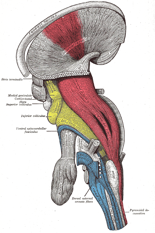 | ||
| Deep dissection of brain-stem. Lateral view. ("pyramidal tract" visible in red, and "pyramidal decussation" labeled at lower right.) | ||
 | ||
| Diagram of the principal fasciculi of the spinal cord. | ||
| Latin | ' | |
| Gray's | subject #185 759 | |
| Part of | ||
| Components | ||
| Artery | ||
| Vein | ||
| BrainInfo/UW | ancil-373 | |
| MeSH | A08.186.854.633 | |
The corticospinal or pyramidal tract is a massive collection of axons that travel between the cerebral cortex of the brain and the spinal cord.
The corticospinal tract contains exclusively motor axons. It actually consists of two separate tracts in the spinal cord: the lateral corticospinal tract and the medial corticospinal tract. An understanding of these tracts leads to an understanding of why for the most part, one side of the body is controlled by the opposite side of the brain.
The motor pathway[]
The corticospinal tract originates in the giant pyramidal neurons (Betz cells) of the motor cortex. The neuronal cell bodies in the motor cortex send long axons to the motor cranial nerve nuclei mainly of the contralateral side of the midbrain (cortico-mesencephalic tract), pons (cortico-pontine tract), medulla oblongata (cortico-bulbar tract); the bulk of these fibers, however, extend all the way down to the spinal cord (corticospinal tract). Most of the cortico-spinal fibers (about 85%) cross over to the contralateral side in the medulla oblongata (pyramidal decussation). Those that do cross in the medulla oblongata travel in the lateral corticospinal tract. The remainder of them (15%) cross over at the level that they exit the spinal cord, and these travel in the medial corticospinal tract. Despite which of these two tracts it travels in, the axon of a neuron which is part of this tract will synapse with another neuron in the ventral horn. This ventral horn neuron is considered a second-order neuron in this pathway, but is not part of the corticospinal tract itself.
There is a precise somatotopic representation of the different body parts in the primary motor cortex, with the leg area located medially (close to the midline), and the head and face area located laterally on the convex side of the cerebral hemisphere (motor homunculus). The arm and hand motor area is the largest and occupies the part of precentral gyrus, located inbetween the leg and face area.
The motor axons move closer together as they travel down through the cerebral white matter, and form part of the posterior limb of the internal capsule.
The motor fibers continue down into the brainstem. The bundle of corticospinal axons is visible as two column-like structures ("pyramids") on the ventral surface of medulla oblongata - this is where the name pyramidal tract comes from.
After the decussation, the axons travel down the spinal cord as the lateral corticospinal tract. Fibers that do not cross over in the medulla oblongata travel down the separate ventral corticospinal tract, and most of them cross over to the contralateral side in the spinal cord, shortly before reaching the lower motor neurons.
The motor neuron cell bodies in the motor cortex, together with their axons that travel down the brain stem and spinal cord, are referred to as upper motor neuron. In the spinal cord, these axons connect (most of them via interneurons, but to a lesser extent also via direct synapses) with the lower motor neurons (LMNs), located in the ventral horn of the spinal cord. In the brain stem, the lower motor neurons are located in the motor cranial nerve nuclei (occulomotor, trochlear, motor nucleus of the trigeminal nerve, abducens, facial, accessory, hypoglossal). The lower motor neuron axons leave the brain stem via motor cranial nerves and the spinal cord via anterior roots of the spinal nerves respectively, end-up at the neuromuscular plate and provide motor innervation for voluntary muscles.
Sensory pathways[]
- Spinothalamic tract
- Spinocerebellar tract
- Visual pathway
- Auditory pathway
- Gustatory pathway
- Olfactory system
- Posterior column pathway
Corticospinal tract damage[]
see upper motor neuron.
Extrapyramidal motor pathways[]
These are motor pathways that lie outside the corticospinal tract and are beyond voluntary control. Their main function is to support voluntary movement and help control posture and muscle tone. See extrapyramidal motor system.
See also[]
References[]
External links[]
| This page uses Creative Commons Licensed content from Wikipedia (view authors). |