Assessment |
Biopsychology |
Comparative |
Cognitive |
Developmental |
Language |
Individual differences |
Personality |
Philosophy |
Social |
Methods |
Statistics |
Clinical |
Educational |
Industrial |
Professional items |
World psychology |
Clinical: Approaches · Group therapy · Techniques · Types of problem · Areas of specialism · Taxonomies · Therapeutic issues · Modes of delivery · Model translation project · Personal experiences ·
| ICD-10 | M89.0, G56.4 | |
|---|---|---|
| ICD-9 | 337.21, 337.22, 354.4, 355.71 | |
| OMIM | {{{OMIM}}} | |
| DiseasesDB | [1] | |
| MedlinePlus | [2] | |
| eMedicine | pmr/123 | |
| MeSH | {{{MeshNumber}}} | |
Complex regional pain syndrome (CRPS) is a chronic condition characterized by severe pain following injury to bone and soft tissue. The International Association for the Study of Pain has divided CRPS into two types based on the presence of nerve lesion following the injury. Type I, also known as Reflex sympathetic dystrophy (RSD), Sudeck's atrophy, Reflex neurovascular dystrophy (RND) or algoneurodystrophy, does not have demonstrable nerve lesions, while type II, also known as causalgia, has evidence of obvious nerve lesions. The cause of these syndromes is currently unknown. Precipitating factors include illness, injury, and psychological stress.[3]
History and nomenclature
The condition currently known as CRPS was originally described by Silas Weir Mitchell during the American Civil War, who named the condition causalgia. In the 1940s, the term reflex sympathetic dystrophy came into use to describe this condition, based on the theory that sympathetic hyperactivity was involved in the pathophysiology (Evans, 1946). Misuse of the terms, as well as doubts about the underlying pathophysiology, led to calls for better nomenclature. In 1993, a special consensus workshop held in Orlando, Florida provided the umbrella term, complex regional pain syndrome, with causalgia and RSD as its subtypes (see Stanton-Hicks et al, 1995).

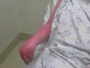
severe CRPS of right arm
Pathophysiology
The pathophysiology of CRPS remains unclear.
Susceptibility
CRPS can strike at any age, but is more common between the ages of 40 and 60. [citation needed] It affects both men and women, but is more frequently seen in women. Although it can occur at any age, the number of CRPS cases among adolescents and young adults is increasing. [citation needed]
Investigators estimate that two to five percent of those with peripheral nerve injury and 12 to 21 percent of those with hemiplegia (paralysis of one side of the body) will suffer from CRPS. [citation needed]
Symptoms
The symptoms of CRPS usually occur near the site of an injury, either major or minor, and usually spreads beyond the original area. It may spread to involve the entire limb and, rarely, the opposite limb. The most common symptom is burning pain. The patient may also experience muscle spasms, local swelling, increased sweating, softening of bones, joint tenderness or stiffness, restricted or painful movement, and changes in the nails and skin.
The pain of CRPS is continuous and may be heightened by emotional stress. Moving or touching the limb is often intolerable. Eventually the joints become stiff from disuse, and the skin, muscles, and bone atrophy. The symptoms of CRPS vary in severity and duration. There are three variants of CRPS, previously thought of as stages. It is now believed that patients with CRPS do not progress through these stages sequentially and/or that these stages are not time limited. Instead, patients are likely to have one of the three following types of disease progression:
- Type one is characterized by severe, burning pain at the site of the injury. Muscle spasm, joint stiffness, restricted mobility, rapid hair and nail growth, and vasospasm (a constriction of the blood vessels) that affects color and temperature of the skin can also occur.
- Type two is characterized by more intense pain. Swelling spreads, hair growth diminishes, nails become cracked, brittle, grooved, and spotty, osteoporosis becomes severe and diffuse, joints thicken, and muscles atrophy.
- Type three is characterized by irreversible changes in the skin and bones, while the pain becomes unyielding and may involve the entire limb. There is marked muscle atrophy, severely limited mobility of the affected area, and flexor tendon contractions (contractions of the muscles and tendons that flex the joints). Occasionally the limb is displaced from its normal position, and marked bone softening is more dispersed
Diagnosis
CRPS types I and II share the common diagnostic crietria shown below.
- Spontaneous pain or allodynia/hyperalgesia is not limited to the territory of a single peripheral nerve, and is disproportionate to the inciting event.
- There is a history of edema, skin blood flow abnormality, or abnormal sweating in the region of the pain since the inciting event.
- No other conditions can account for the degree of pain and dysfunction.
The two types differ only in the nature of the inciting event. Type I CRPS develops following an initiating noxious event that may or may not have been traumatic, while type II CRPS develops after a nerve injury.
No specific test is available for CRPS, which is diagnosed primarily through observation of the symptoms. However, thermography, sweat testing, x-rays, electrodiagnostics, and sympathetic blocks can be used to build up a picture of the disorder. Diagnosis is complicated by the fact that some patients improve without treatment. A delay in diagnosis and/or treatment for this syndrome can result in severe physical and psychological problems. Early recognition and prompt treatment provide the greatest opportunity for recovery.
Thermography
Thermography is a diagnostic technique for measuring blood flow by determining the variations in heat emitted from the body. A color-coded "thermogram" of a person in pain often shows an altered blood supply to the painful area, appearing as a different shade (abnormally pale or violet) than the surrounding areas of the corresponding part on the other side of the body. A difference of 1.0°C between two symmetrical body parts is considered significant, especially if a large number of asymmetrical skin temperature sites are present. [citation needed] The affected limb may be warmer or cooler than the unaffected limb.
Sweat testing
Abnormal sweating can be detected by several tests. A powder that changes color when exposed to sweat can be applied to the limbs; however, this method does not allow for quantification of sweating. Two quantitative tests that may be used are the resting sweat output test and the quantitative sudomotor axon reflex test. These quantitative sweat tests have been shown to correlate with clinical signs of CRPS (Sandroni, 1998).
Radiography
Patchy osteoporosis, which may be due to disuse of the affected extremity, can be detected on X-ray as early as 2 weeks after the onset of CRPS. Bone scan of the affected limb may detect these changes even sooner. Bone densitometry can also be used to detect changes in bone mineral density. It can also be used to monitor the results of treatment, as bone densitometry paramters improve with treatment.
Electrodiagnostic testing
The nerve injury that characterizes type II CRPS can be detected by electromyography. In contrast to peripheral mononeuropathy, the symptoms of type 2 CRPS extend beyond the distribution of the affected peripheral nerve.
Treatment
Physicians use a variety of drugs to treat CRPS, including antidepressants, corticosteroids, vasodilators, gabapentin, pregabalin,and alpha- or beta-adrenergic-blocking compounds. Elevation of the extremity and physical therapy are also used to treat CRPS. Injection of a local anesthetic, such as lidocaine, is often the first step in treatment. Injections are repeated as needed. However, early intervention with non-invasive management may be preferred to repeated nerve blockade. TENS (transcutaneous electrical nerve stimulation), a procedure in which brief pulses of electricity are applied to nerve endings under the skin, has helped some patients in relieving chronic pain. Neurostimulation (spinal cord stimulators) may also be surgically implanted to reduce the pain by directly stimulating the spinal cord. These devices place electrodes either in the epidural space (space above the spinal cord) or directly over nerves located outside the central nervous system. Implantable drug pumps may also be used to deliver pain medication directly to the cerebrospinal fluid which allows the use of powerful opioids to be used in a much smaller dose than when taken orally. Ketamine infusion to treat CRPS has been described (Correll et al., 2004). Prednisolone (a corticosteroid) has been shown to be superior to piroxicam in the treatment of reflex sympathetic dystrophy.[1]
Surgical, chemical, or radiofrequency sympathectomy — interruption of the affected portion of the sympathetic nervous system — can be used as a last resort in patients with impending tissue loss, edema, recurrent infection, or ischemic necrosis (Stanton-Hicks et al, 1998). However, there is little evidence that these permanent interventions alter the pain symptoms of these devastated patients.
Physical therapy is the most important part of treatment, though it should be noted that many patients are incapable of participating in physical therapy due to muscular and bone problems. People struggling with CRPS often develop guarding behaviors where they avoid using or touching the affected limb. Unfortunately, inactivity can exacerbate the disease and perpetuate the pain cycle. Physical therapy works best for most patients, especially goal-directed therapy, where the patient begins from an initial point, regardless of how minimal, and then endeavors to increase activity each week. Therapy should be directed at facilitating the patient to engage in physical therapy, movement and stimulation of the affected areas. Some treating physicians have even initiated physical therapy under light general anesthesia, in an attempt to remobilze the extremity. While the unpredictability of this illness often causes a frustrating pattern of progress and regress, it is essential to continue to try to increase and normalize physicial activity. It hurts loads though.
EEG Biofeedback and Feldenkrais can also be important modalities of treatment.
Prognosis
Good progress can be made in treating CRPS if treatment is begun early, ideally within 3 months of the first symptoms. Early treatment often results in remission. If treatment is delayed, however, the disorder can quickly spread to the entire limb and changes in bone and muscle may become irreversible. In 50 percent of CRPS cases, pain persists longer than 6 months and sometimes for years. [citation needed] In teens and younger patients with CRPS, the prognosis is excellent. Even without invasive therapy, upwards of 75% of children have full recovery with virtually 100% of the patients have marked improvement.
Similar disorders
CRPS has characteristics similar to those of other disorders, such as shoulder-hand syndrome, which sometimes occurs after a heart attack and is marked by pain and stiffness in the arm and shoulder; Sudeck syndrome, which is prevalent in older people and women and is characterized by bone changes and muscular atrophy, but is not always associated with trauma; and Steinbrocker syndrome, which includes symptoms such as gradual stiffness, discomfort, and weakness in the shoulder and hand. Erythromelalgia also shares many components of CRPS (burning pain, redness, tempurature hypersensative, autonomic dysfunction, vasospasm)they both involve small fiber sensory neurosympathetic components. Interestingly Erythromelalgia involves a lack of sweating, whereas CRPS often involves increased sweating. Subvariations of both exist.
Current research
The National Institute of Neurological Disorders and Stroke (NINDS), a part of the National Institutes of Health (NIH), supports and conducts research on the brain and central nervous system, including research relevant to RSDS, through grants to major medical institutions across the country. NINDS-supported scientists are working to develop effective treatments for neurological conditions and, ultimately, to find ways of preventing them.Investigators are studying new approaches to treat RSDS and intervene more aggressively after traumatic injury to lower the patient's chances of developing the disorder. In addition, NINDS-supported scientists are studying how signals of the sympathetic nervous system cause pain in RSDS patients. Using a technique called microneurography, these investigators are able to record and measure neural activity in single nerve fibers of affected patients. By testing various hypotheses, these researchers hope to discover the unique mechanism that causes the spontaneous pain of RSDS and that discovery may lead to new ways of blocking pain.Other studies to overcome chronic pain syndromes are discussed in the pamphlet "Chronic Pain: Hope Through Research," published by the NINDS.
CRPS in animals
CRPS has also been described in non-human animals (Bergadano et al, 2006).
External links
- RSD Support Group
- Forgrace.org
- RSDhope.org
- RSDS Fact Sheet
- NINDS Complex Regional
- Pain Syndrome Information Page
- Canadian RSD
- RSD Information Personal Experience
References
- Bergadano A, Moens Y, Schatzmann U (2006). Continuous extradural analgesia in a cow with complex regional pain syndrome. Vet Anaesth Analg 33 (3): 189-92. PMID 16634945.
- Birklein F (2005). Complex regional pain syndrome. J Neurol 252 (2): 131-8. PMID 15729516.
- Correll GE, Maleki J, Gracely EJ, Muir JJ, Harbut RE (2004). Subanesthetic ketamine infusion therapy: a retrospective analysis of a novel therapeutic approach to complex regional pain syndrome. Pain Med 5 (3): 263-75. PMID 15367304.
- Evans JA (1946). Reflex sympathetic dystrophy. Surg Clin North America 26: 780-790.
Lee,B.H.; Scharff,L.; Sethna,N.F.; McCarthy,C.F.; Scott-Sutherland,J.; Shea,A.M.; Sullivan,P.; Meier,P.; Zurakowski,D.; Masek,B.J.; Berde,C.B. Physical therapy and cognitive-behavioral treatment for complex regional pain syndromes Source: J Pediatr, 2002, 141, 1, 135-40
- Sandroni P, Low PA, Ferrer T, Opfer-Gehrking TL, Willner CL, Wilson PR (1998). Complex regional pain syndrome I (CRPS I): prospective study and laboratory evaluation. Clin J Pain 14 (4): 282-9. PMID 9874005.
- Stanton-Hicks M, Janig W, Hassenbusch S, Haddox JD, Boas R, Wilson P (1995). Reflex sympathetic dystrophy: changing concepts and taxonomy. Pain 63 (1): 127-33. PMID 8577483.
- Stanton-Hicks M, Baron R, Boas R, Gordh T, Harden N, Hendler N, Koltzenburg M, Raj P, Wilder R (1998). Complex Regional Pain Syndromes: guidelines for therapy. Clin J Pain 14 (2): 155-66. PMID 9647459.
Wilder,R.T.; Berde,C.B.; Wolohan,M.; Vieyra,M.A.; Masek,B.J.; Micheli,L.J. |Title=Reflex sympathetic dystrophy in children. Clinical characteristics and follow-up of seventy patients | Journal=Journal of Bone & Joint Surgery - American | Year=1992 | Pages=910-19 | volume=74 | Issue=6
- de:Komplexes regionales Schmerzsyndrom
- fr:Algoneurodystrophie
- nl:Causalgie
- pl:Zespół algodystroficzny
| This page uses Creative Commons Licensed content from Wikipedia (view authors). |
- ↑ Kalita J, Vajpayee A, Misra UK. (2006). Comparison of prednisolone with piroxicam in complex regional pain syndrome following stroke: a randomized controlled trial.. QJM 99 (2): 89–95.