Assessment |
Biopsychology |
Comparative |
Cognitive |
Developmental |
Language |
Individual differences |
Personality |
Philosophy |
Social |
Methods |
Statistics |
Clinical |
Educational |
Industrial |
Professional items |
World psychology |
Biological: Behavioural genetics · Evolutionary psychology · Neuroanatomy · Neurochemistry · Neuroendocrinology · Neuroscience · Psychoneuroimmunology · Physiological Psychology · Psychopharmacology (Index, Outline)
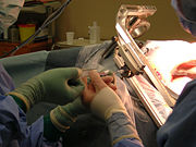
Brain biopsy
A biopsy (in Greek: bios = life and opsy = look/appearance) is a medical test involving the removal of cells or tissues for examination. The tissue is generally examined under a microscope by a pathologist, and can also be analyzed chemically (for example, using PCR or gas chromatography techniques). When only a sample of tissue is removed, the procedure is called an incisional biopsy or core biopsy. When an entire lump or suspicious area is removed, the procedure is called an excisional biopsy. When a sample of tissue or fluid is removed with a needle, the procedure is called a needle aspiration biopsy.
After the biopsy is performed, the sample of tissue that was removed from the patient is sent to the pathology laboratory. A pathologist is a physician who specializes in diagnosing diseases (such as cancer) by examining tissue under a microscope. When the laboratory receives the biopsy sample, the tissue is processed and an extremely thin slice of tissue is removed from the sample and attached to a glass slide. Any remaining tissue is saved for use in later studies, if required. The slide with the tissue attached is treated with dyes that stain the tissue, which allows the individual cells in the tissue to be seen more clearly. The slide is then given to the pathologist, who examines the tissue under a microscope, looking for any abnormal findings. The pathologist then prepares a report that lists any abnormal or important findings from the biopsy. This report is sent to the physician who originally performed the biopsy on the patient.
In cancer[]
Pathologic examination of a biopsy can determine whether a lesion is benign or [Malignant_tumor and can help differentiate between different types of cancer. In contrast to a biopsy that merely samples a lesion, a larger excisional specimen called a resection may come to a pathologist, typically from a surgeon attempting to eradicate a known lesion from a patient. For example, a pathologist would examine a mastectomy specimen, even if a previous nonexcisional breast biopsy had already established the diagnosis of breast cancer. Examination of the full mastectomy specimen would confirm the exact nature of the cancer (subclassification of tumor and histologic "grading") and reveal the extent of its spread (pathologic "staging").
In an excisional biopsy, the margins of the specimen may be carefully examined to see if the disease has spread beyond the area biopsied. "Clear margins," or "negative margins," means that no disease was found at the edges of the biopsy specimen. "Positive margins" means that disease was found, and additional treatment may be needed.
Other[]
Biopsy specimens are often taken from part of a lesion when the cause of a disease is uncertain or its extent or exact character is in doubt. Vasculitis, for instance, is usually diagnosed on biopsy.
A testicular biopsy is used for evaluating the fertility of men and find out the cause of a possible infertility, e.g. when sperm quality is low, but hormone levels still are within normal ranges. [1]
External links[]
- MyBiopsy.org - Information about biopsy results for patients. This site is created by pathologists, the physicians who diagnose cancer and other diseases by looking at biopsies under a microscope.
- RadiologyInfo - The radiology information resource for patients: Biopsy
References[]
Template:Pathology-stub
| This page uses Creative Commons Licensed content from Wikipedia (view authors). |